
System and method for lung visualization using ultasound

Active Publication Date: 2015-03-12
TYCO HEALTHCARE GRP LP
View PDF9 Cites 55 Cited by
  • Summary
  • Abstract
  • Description
  • Claims
  • Application Information

AI Technical Summary

Benefits of technology

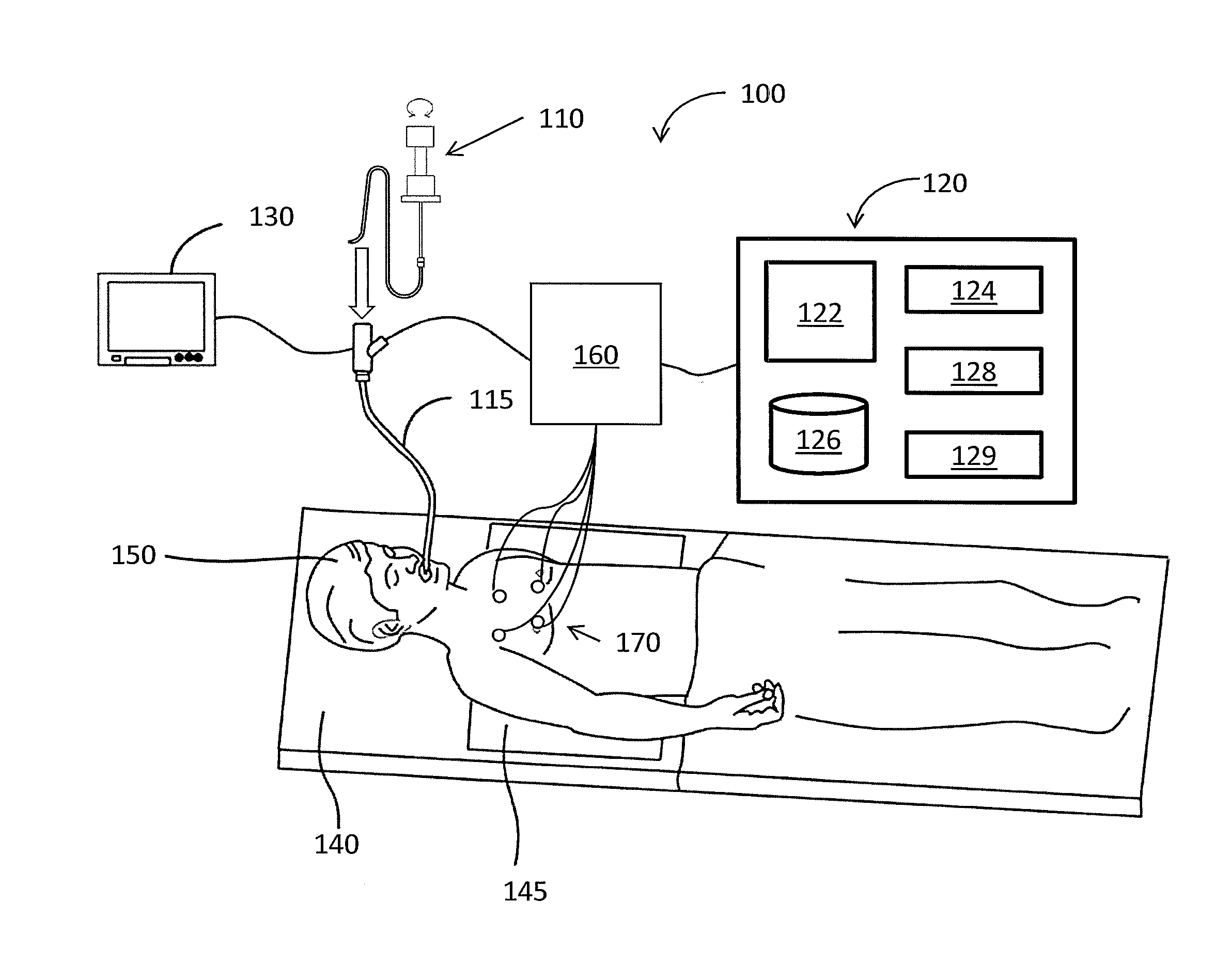
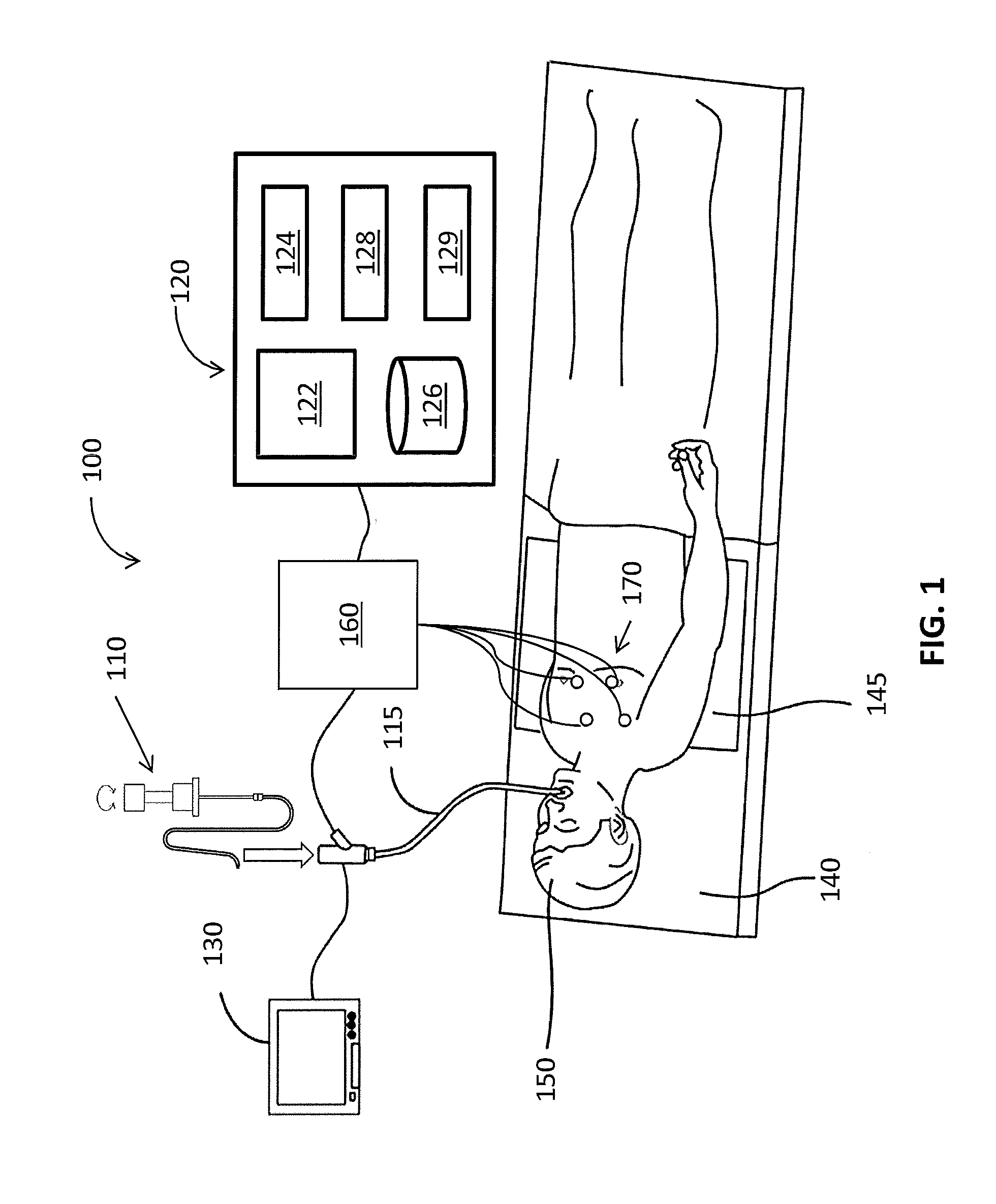
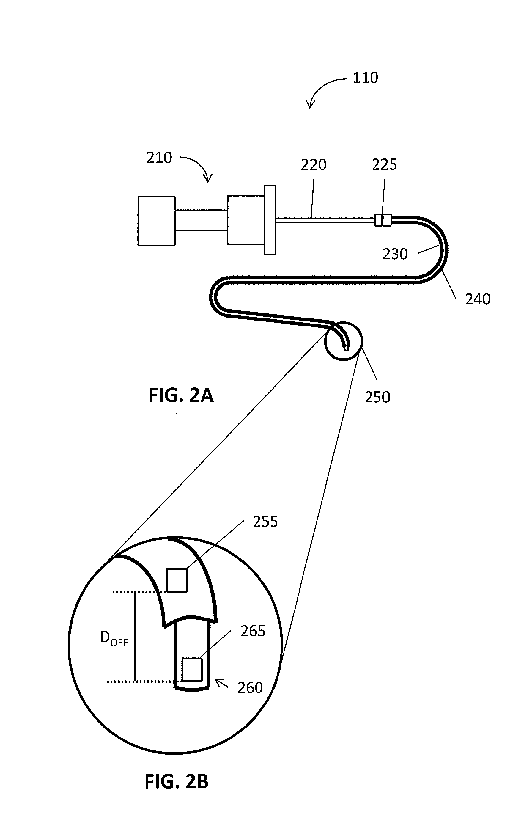
The present patent describes a system for interrogating a lung in the US based on a 3D model and a pathway plan. The system includes an EM board, an EWC, an EM sensor, a US transducer, and a processor. The EM sensor senses the EM field generated by the EM board and the US transducer generates US waves that are reflected from the lung. The processor processes the sensed EM field to synchronize its location in the 3D model and generate US images that can be integrated with the 3D model. The system also includes a display device to display the integrated 3D model and US images, with a status based on the location of the EM sensor. The generated images have a finer resolution than the 3D model. The system also includes reference sensors and a tracking device to identify the location of the EM sensor and compensate for patient's breathing. The processor can also determine the density, size, and shrinkage of the target during treatment. The generated images show outside of the lung and the offset between the EM sensor and the US transducer is determined. The technical effects of the system include improved accuracy and precision in lung interrogation and treatment.

Problems solved by technology

However, images generated by these non-invasive imaging technologies have been unable to provide a resolution sufficient to identify features such locations of nerves that run parallel to the luminal network.
That increases the number of exposures of harmful X-rays or substances to the patient and costs of treatments.

Method used


Image

  • System and method for lung visualization using ultasound
  • System and method for lung visualization using ultasound
  • System and method for lung visualization using ultasound

Examples


Embodiment Construction

[0032]The present disclosure is related to systems and methods for visualizing the luminal network of a lung using ultrasound (US) imaging technologies which provide a sufficient resolution to identify and locate a target for diagnostic, navigation, and treatment purposes. US imaging, particularly in conjunction with non-invasive imaging can provide a greater resolution and enable luminal network mapping and target identification. Further, additional clarity is provided with respect to tissue adjacent identified targets which can result in different treatment options being considered to avoid adversely affecting the adjacent tissue. Still further, the use of US imaging in conjunction with treatment can provide detailed imaging for post treatment analysis and identification of sufficiency of treatment. Although the present disclosure will be described in terms of specific illustrative embodiments, it will be readily apparent to those skilled in this art that various modifications, re...


PUM


Abstract

A system for ultrasound interrogation of a lung includes a memory, an electromagnetic (EM) board, an extended working channel (EWC), an EM sensor, a US transducer, and a processor. The memory stores a three dimensional (3D) model, a pathway plan for navigating a luminal network. An EM board generates an EM field. The EWC is configured to navigate the luminal network of a patient toward a target following the pathway plan and the EM sensor extends distally from the EWC and senses the EM field. The US transducer extends distally from a distal end of the EWC and generates US waves and receives US waves reflected from the luminal network and the processor processes the sensed EM field to synchronize a location of the EM sensor in the 3D model, to process the reflected US waves to generate images, or to integrate the generated images with the 3D model.

Description

BACKGROUND[0001]1. Technical Field[0002]The present disclosure relates to systems and methods for visualizing a lung using ultrasound imaging techniques. More particularly, the present disclosure relates to systems and methods that augment images of a luminal networks obtained by other imaging modality with ultrasound images.[0003]2. Discussion of Related Art[0004]Standard of care for lung diseases, such as asthma, chronic obstructive pulmonary disease (COPD), and chronic obstructive lung disease (COLD), or for lung-related diseases has been focused largely on medical and / or drug management which are highly invasive to patients in general. For example, it has been reported for decades that lung denervation via localized and invasive means (e.g., surgery) may provide therapeutic benefit for asthma or emphysema.[0005]Electromagnetic navigation (EMN) has helped expand the possibilities of treatment of luminal networks such as the lungs. EMN relies on non-invasive imaging technologies, ...

Claims


Application Information

Patent Timeline
IPC IPC(8): A61B5/06A61B8/12A61B8/08
CPCA61B5/062A61B8/12A61B8/5207A61B1/267A61B8/08A61B8/463A61B8/467A61B8/5261A61B5/113A61B5/7207A61B8/4254A61B8/5238
Inventor BRANNAN, JOSEPH D.DICKHANS, WILLIAM J.LADTKOW, CASEY M.PETERSON, DARION R.
Owner TYCO HEALTHCARE GRP LP
Learn More