Elastography-based assessment of cryoablation

a cryoablation and elastography technology, applied in the field of tissue cryoablation, can solve the problems of inability to monitor cryoablation in real time with conventional ultrasound, inability to monitor cryoablation boundaries with standard b-mode ultrasound, and inability to provide good contrast of the region

Inactive Publication Date: 2015-03-26
THE UNIV OF BRITISH COLUMBIA
View PDF1 Cites 8 Cited by
  • Summary
  • Abstract
  • Description
  • Claims
  • Application Information

AI Technical Summary

Problems solved by technology

It is well known that real-time monitoring of cryoablation with conventional ultrasound is not possible because during the freezing cycle of the cryoablation process, an ice ball is found that overlaps with the tumour region to be ablated.
Alternatively, B-Mode ultrasound images are used in ultrasound guided cryoablation to visualize the frozen zone after the complete thawing, but these images do not provide good contrast of the region (Onik, G M, G Reyes, J K Cohen, and B Porterfield , “Ultrasound characteristics of renal cryosurgery.” Urology, 42, 212-215, 1993).
Therefore, the boundaries of the cryoablated areas cannot be monitored with standard B-mode ultrasound as the ablation proceeds.
For assessment of a complete cryoablation freeze-thaw cycle, standard B-mode ultrasound is not accurate enough.

Method used


Image

  • Elastography-based assessment of cryoablation
  • Elastography-based assessment of cryoablation
  • Elastography-based assessment of cryoablation

Examples


Embodiment Construction

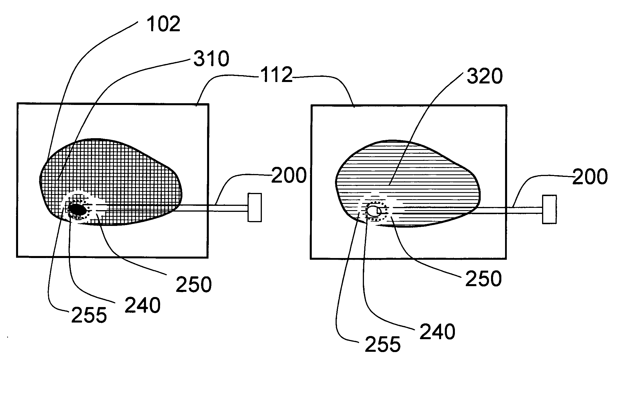
[0020]We propose to use the elasticity of tissue, measured with ultrasound-based elastography, to determine whether a region of tissue has been properly cryoablated. Our description will be with reference to a cryoablation system for the prostate, but it is understood that this reference to the prostate is in no way limiting and that applications to other organs will be similar and easily adapted to by someone skilled in the art.

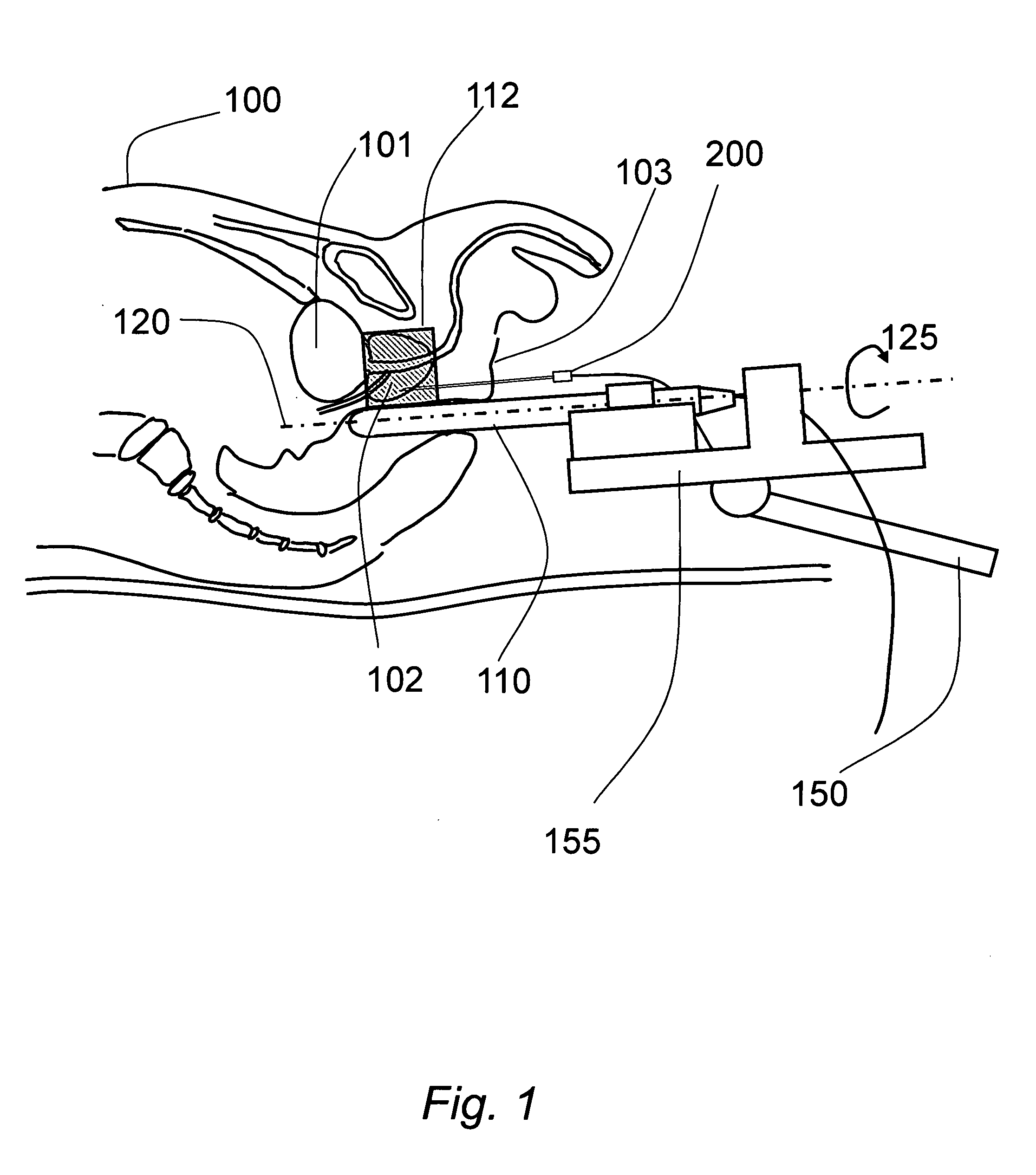
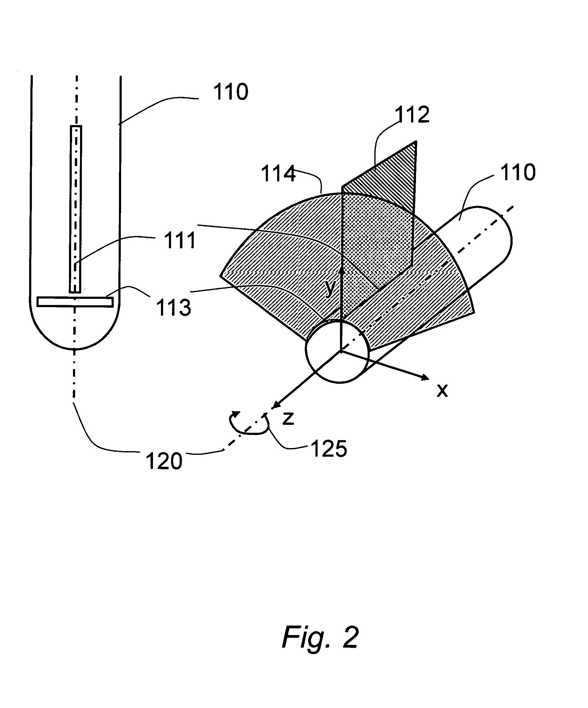
[0021]Aspects of the invention are described with reference to FIGS. 1 through 5 and FIG. 8 as applied to prostate ablation. For a cryoablation procedure, the patient 100 is being imaged by an ultrasound transducer 110 that images the prostate 102 as well as some of the periprostatic region including part of the bladder 101. The transducer is connected to a brachytherapy stabilizer 150 and stepper 155, as exemplified by the CIVCO MicroTouch stabilizer with the EXII Brachytherapy Stepper. The transducer, shown in FIG. 2, is a brachytherapy transducer having a...


PUM


Abstract

A method of monitoring the cryoablation of a target volume of tissue with ultrasound elastography, the method comprising acquiring a first elastography image encompassing said target volume of tissue, performing at least one cycle of freezing and thawing of tissue encompassed in said target volume, acquiring a second elastography image encompassing said target volume, and comparing said first and said second elastography images over said target volume. The elastography provides either relative or quantitative measurements of tissue elasticity. The elastography maps of tissue elasticity, before and after cryoablation of one region, can guide the cryoablation of another region. The use of elastography provided feedback to the operator to achieve effective treatment with cryoblation over a planned target.

Description

FIELD OF THE INVENTION[0001]The present invention relates to tissue cryoablation and, in particular, to a method of assessing the result of cryoablation for quality assurance and treatment re-planning.BACKGROUND OF THE INVENTION[0002]The basis of medical imaging is the measurement of a property of tissue that varies with tissue composition. Medical images are formed by displaying intensities as a function of these properties measured at various locations in the body. Mechanical properties of tissue are important indicators of disease potential. Indeed, palpation techniques are commonly used by medical doctors to determine the potential for disease, for example, stiffer tissue regions that feel harder can indicate the presence of cancer. This is the basis for a number of clinical examinations such as the digital rectal examination for prostate cancer. A change in the mechanical properties of tissue can also be an indicator of the success or failure of therapy.[0003]Elastography is a ...

Claims


Application Information

Patent Timeline
Patent Type & Authority Applications(United States)
IPC IPC(8): A61B8/08A61B18/02
CPCA61B8/0858A61B2018/00642A61B2018/00577A61B18/02A61B2018/0293A61B8/085A61B8/12A61B8/485A61B2090/3782
Inventor SALCUDEAN, SEPTIMIUSCHNEIDER, CAITLINDALL'ALBA, DIEGOROHLING, ROBERTNGUAN, CHRISTOPHER
Owner THE UNIV OF BRITISH COLUMBIA
Why Eureka
Try Eureka