Methods for Tissue Passivation

a tissue and passivation technology, applied in the field of tissue passivation, can solve the problems of affecting the quality of life of patients, the inability to achieve effective prevention methods of capsular contracture, and the deformation or rupture of the capsule, so as to prevent or reduce the stenosis, the effect of preventing or reducing the stenosis

Active Publication Date: 2015-05-14
THE GENERAL HOSPITAL CORP
View PDF12 Cites 14 Cited by
  • Summary
  • Abstract
  • Description
  • Claims
  • Application Information

AI Technical Summary

Benefits of technology

[0056]In yet another aspect, the invention provides a method of preventing or reducing stenosis in a subject, the method comprising the steps of: implanting a graft comprising vein into an artery, wherein the implanting of the graft replaces and or b

Problems solved by technology

In capsular contracture, these processes go awry, resulting in dense, linear bundles of collagen fibers that surround the affected implant.
Direct pressure from a maturing capsule may deform or rupture the implant, in addition to distorting the overlying skin and soft tissue.
The condition may be painful and debilitating as well as aesthetically inferior.
Despite decades of research, effective methods of prevention of capsular contracture remain elusive3.
Saphenous vein grafts (SVGs) used for coronary arte

Method used


Image

  • Methods for Tissue Passivation
  • Methods for Tissue Passivation
  • Methods for Tissue Passivation

Examples


example 1

Tissue Surface Passivation in a Rabbit Model for Capsular Contraction

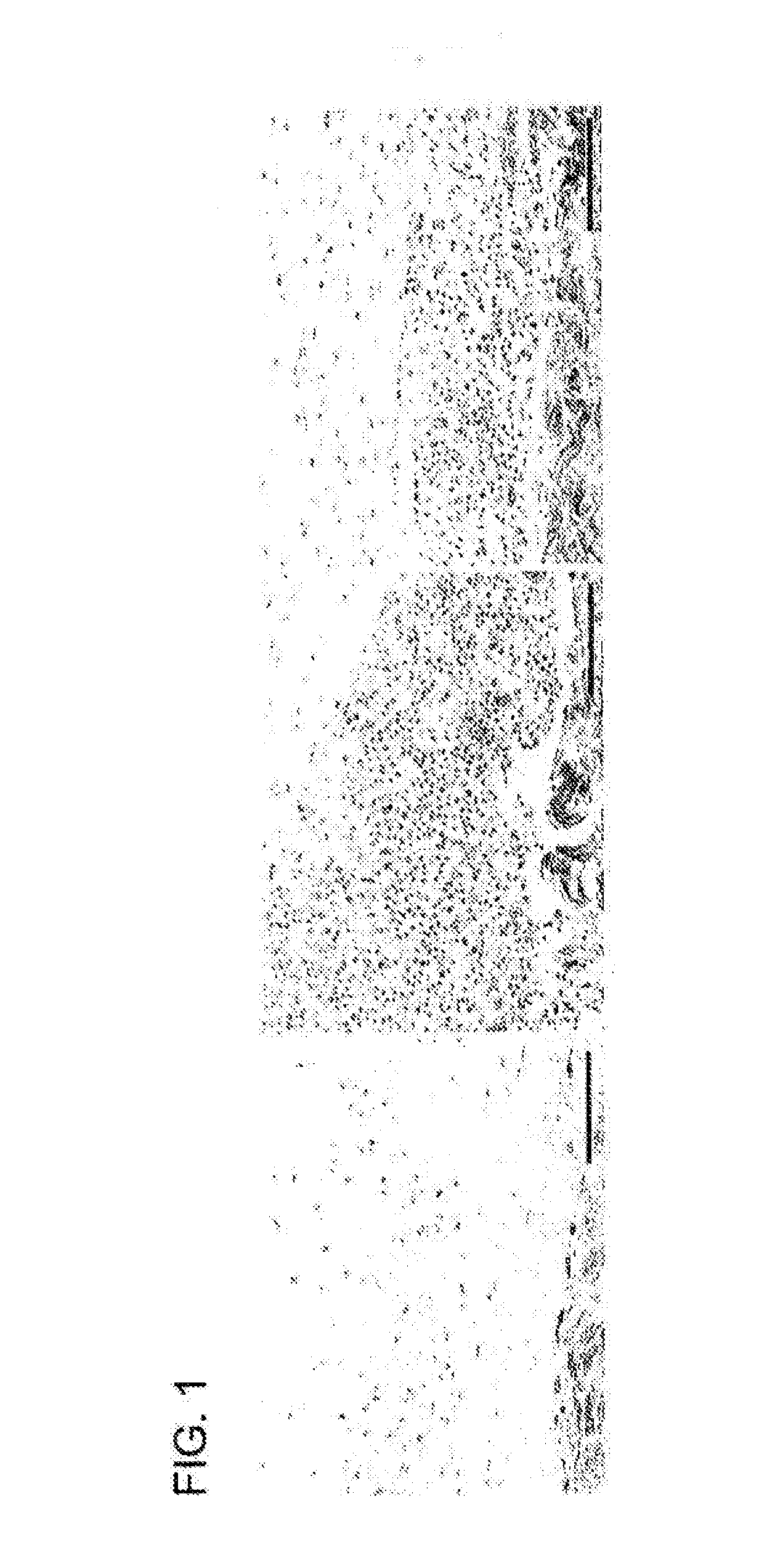
[0121]A pilot experiment in a rabbit model for capsular contraction was developed. Three sub-cutaneous pockets were made by surgical incision on the upper rabbit flank. One pocket was left untreated and surgically closed (No fibrin glue). Of the remaining two, one was treated with Fibrin Glue only, and one received Fibrin Glue+RB (0.1% Rose Bengal)+532 nm light (3 min at 350 mW). Fibrin glue is known to produce a fibrotic reaction in this model. After two weeks all three pockets were surgically re-accessed and inspected. Tissue harvested at two weeks demonstrated an aggressive inflammatory response, with a significant infiltrate within the implant pockets instilled with fibrin glue (FIG. 1). The pockets treated with PTP prior to fibrin glue application, had less infiltrate. These cells also appear contained under layers of presumably, cross-linked collagen. A biodegradation assay was also performed. Pockets were tr...

example 2

Tissue Surface Passivation in a Pre-Clinical Model of Capsular Contracture Following Prosthetic Implant Placement

[0123]The effect of Photochemical Tissue Passivation in a pre-clinical model of capsular contracture following prosthetic implant placement was evaluated. Under general anesthesia, Nine New Zealand white rabbits received three, six cc smooth saline implants placed in the dorsal sub-panniculus soft tissue. Each Rabbit received one control implant and two experimental implants.

[0124]Briefly, the subpanniculus carnosus plane in the dorsum region was dissected and a single customized six cc saline smooth implant with pressure port valve (Allergan Inc., CA, USA) was placed. A total of three implants were placed on the dorsum of each rabbit. The control group consisted of an implant with no Fibrin Glue and no PTP treatment. Before implant placement, the pocket site of the PTP experimental group was treated with 2 ml 0.1% Rose Bengal in a phosphate buffered saline, and was then ...

example 3

Photochemical Tissue Passivation for Prevention of Vein Graft Intimal Hyperplasia

[0131]Vein grafts are frequently used for Coronary Artery Bypass Grafting yet suffer poor long-term patency rates compared to arterial grafts due to accelerated atherosclerosis. This begins as intimal hyperplasia (IH), which is a consequence of intimal injury resulting from surgical trauma and excessive dilation of the vein graft as it is exposed to arterial pressures. Limiting this stretch reduces the degree of IH. To date, this has only been achieved with external sheaths. Photochemical Tissue Passivation (PTP) was conducted to determine whether this procedure could improve the long-term patency rates of venous grafts.

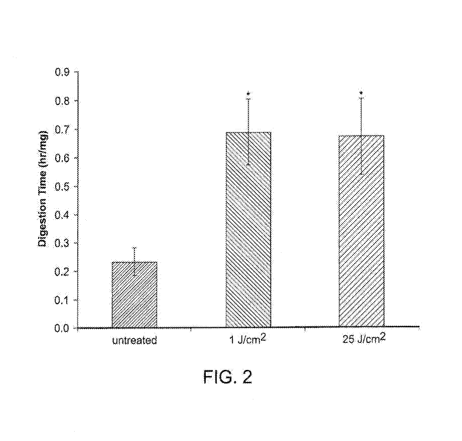
[0132]Porcine jugular veins were used to evaluate the effect of PTP on the elasticity of venous tissues. Veins harvested from five pigs were divided in half. One segment served as control, the other was treated (PTP) with 0.1% Rose Bengal and a 532 nm laser (delivering 25 J / cm2). Veins w...


PUM

PropertyMeasurementUnit
Areaaaaaaaaaaa
Structureaaaaaaaaaa
Adhesivityaaaaaaaaaa

Abstract

Methods of tissue passivation are described herein for use in preserving normal tissue architecture, reducing post-surgical inflammation, and reducing or preventing the development of pathogenic collagen bundles and adhesions following surgical procedures. Passivated tissues prepared in accordance with these methods are useful in a variety of therapies including, e.g., cardiac bypass surgery, hemodialysis, etc.

Description

CROSS-REFERENCE TO RELATED APPLICATIONS[0001]This application is a continuation-in-part of international application Ser. No. PCT / US2013 / 051333, filed on Jul. 19, 2013 and published in English as WO 2014 / 015274 on Jan. 23, 2014, which claims the benefit of U.S. provisional application Ser. Nos. 61 / 674,235, filed on Jul. 20, 2012, 61 / 784,708, filed on Mar. 14, 2013 and 61 / 847,794, filed on Jul. 18, 2013. The entire disclosure of each of the aforementioned patent applications is incorporated herein by reference.[0002]This application may also contain subject matter that is related to that disclosed in PCT / US2014 / 047146, filed Jul. 19, 2014, the entire disclosure of which is incorporated herein by referenceBACKGROUND OF THE INVENTION[0003]Capsular contracture is one of the most common causes of reoperation following implantation3,6. The etiology of capsular contracture has been studied for many years3. Potential etiologies include hematoma, hypertrophic scar and, microbacterial infecti...

Claims


Application Information

Patent Timeline
IPC IPC(8): A61F2/06A61L27/50A61K41/00A61N5/06A61L27/54
CPCA61F2/06A61N5/062A61N5/0601A61L27/54A61K41/0057A61L2430/34A61L2300/418A61L2300/442A61L2400/18A61L2430/04A61L2430/22A61L27/505A61L27/50A61L27/3604A61L27/3633A61L2300/424A61L31/005A61L31/14A61L31/16A61N5/0603A61N5/0624
Inventor AUSTEN, JR., WILLIAM G.MCCORMACK, MICHAELREDMOND, ROBERT W.KOCHEVAR, IRENE E.
Owner THE GENERAL HOSPITAL CORP
Why Eureka
Try Eureka