Image processing apparatus, imaging system, and image processing system

Inactive Publication Date: 2015-06-04
CANON KK
View PDF1 Cites 19 Cited by
  • Summary
  • Abstract
  • Description
  • Claims
  • Application Information

AI Technical Summary

Benefits of technology

[0008]According to the method described in PTL 1, an image focused over the entire range and with little blur can be obtained, and thus a merit is provided that it can be comprehended the condition of the object as a whole with only one image. However, although such deep-focus image is useful for rough observation of th

Problems solved by technology

However, although such deep-focus image is useful for rough observation of the object as a whole, it is not suitable for detailed observation of a part of the object or comprehension of the three-dim

Method used


Image

  • Image processing apparatus, imaging system, and image processing system
  • Image processing apparatus, imaging system, and image processing system
  • Image processing apparatus, imaging system, and image processing system

Examples


first embodiment

System Configuration

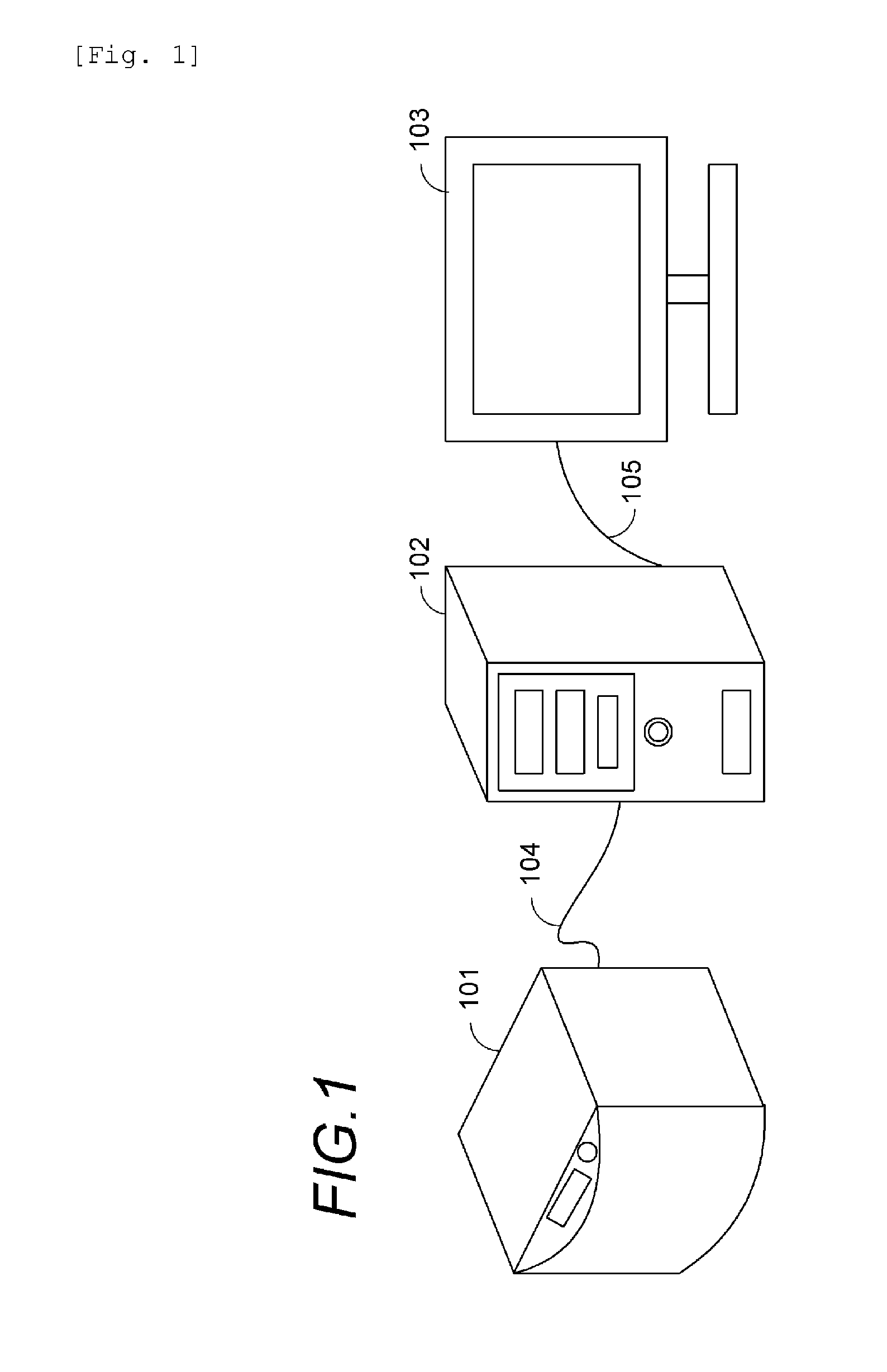
[0036]FIG. 1 is an overall view showing a layout of apparatuses in an imaging system according to a first embodiment of the invention.

[0037]The imaging system according to the first embodiment is composed of an imaging apparatus 101, an image processing apparatus 102, and a display device 103, and is a system with a function to acquire and display a two-dimensional image of a specimen (object) as an object to be imaged. The imaging apparatus 101 and the image processing apparatus 102 are connected to each other with a dedicated or general-purpose I / F cable 104. The image processing apparatus 102 and the display device 103 are connected to each other with a general-purpose I / F cable 105.

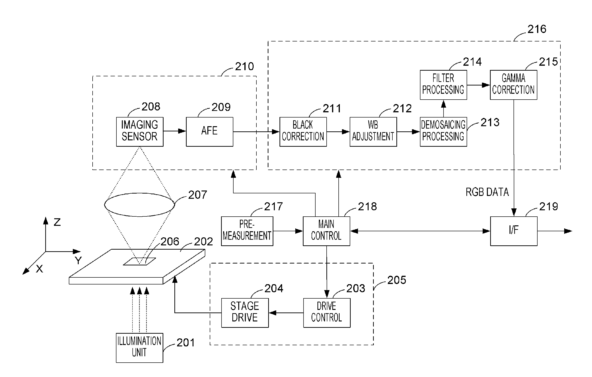
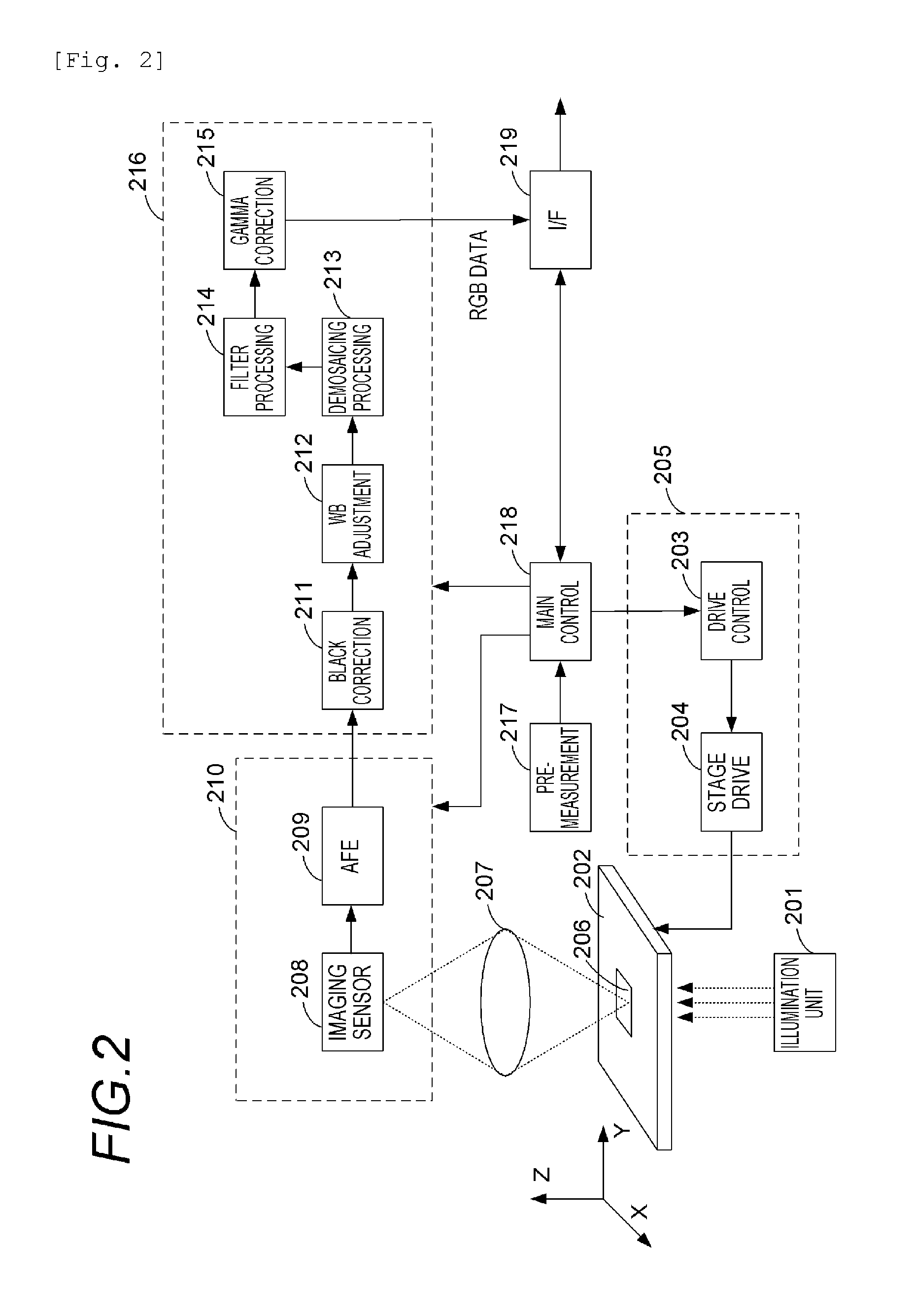
[0038]The imaging apparatus 101 is a microscope apparatus (a virtual slide apparatus) having a function of acquiring a plurality of two-dimensional images at different focal positions in an optical axis direction and outputting digital images. The acquisition of the two-dimensional...

second embodiment

[0103]A second embodiment of this invention will be described. The description of the first embodiment has been made on the configuration for realizing the observation method in which the depth of field is varied while the focal position is kept fixed is described. However, in this second embodiment, a configuration for realizing an observation method in which the focal position is varied while the depth of field is kept fixed.

[0104](System Configuration)

[0105]FIG. 10 is an overall view illustrating a layout of apparatuses in an image processing system according to the second embodiment.

[0106]The image processing system according to this second embodiment is composed of an image server 1201, an image processing apparatus 102, and a display device 103. The second embodiment is different from the first embodiment in that whereas the image processing apparatus 102 in the first embodiment acquires an image from the imaging apparatus 101, the image processing apparatus 102 in the second ...

third embodiment

[0134]A third embodiment of this invention will be described. One of characteristics of the image processing apparatus 102 according to the embodiment resides in that a combined image can be obtained by selectively performing the combine methods described in the embodiments above. Another characteristic of the image processing apparatus 102 according to the third embodiment is that the display method described in the embodiments above and other display method to be described later are selectively performed. Description will be made focusing on these points.

[0135]FIG. 15 is flowchart illustrating a flow of image acquisition according to this third embodiment. In step S1701, the image processing apparatus 102 allows the user to select an image acquisition mode. The image can be acquired by selecting any of a local storage device in the image processing apparatus 102, the image server 1201, and the imaging apparatus 101 as the source of acquisition of the image.

[0136]When the local sto...


PUM


Abstract

An image processing apparatus includes: an image acquisition unit configured to acquire layer images obtained by imaging different positions of an object by using a microscope; an image generation unit for generating a plurality of observation images from the layer images. The image generation unit generates the observation images by performing combine processing for focus-stacking two or more layer images selected from among the layer images to generate a single observation image, for plural times.

Description

TECHNICAL FIELD[0001]This invention relates to an image processing apparatus, an imaging system, and an image processing system, and in particular to a technique for assisting observation of an object with the use of a digital image.BACKGROUND ART[0002]Recently, a virtual slide system attracts attention in the field of pathology, as a successor to an optical microscope which is currently used as a tool for pathological diagnosis. The virtual slide system enables pathological diagnosis to be performed on a display by imaging a specimen to be observed placed on a slide and digitizing the image. The digitization of pathological diagnosis images with the virtual slide system makes it possible to handle conventional optical microscope images of specimens as digital data. It is expected this will bring about various merits, such as more rapid remote diagnosis, provision of information to patients through digital images, sharing of data of rare cases, and more efficient education and train...

Claims


Application Information

Patent Timeline
IPC IPC(8): G02B21/36H04N13/395
CPCG02B21/368G02B21/367H04N13/395G06T1/0007
Inventor SATO, KAZUYUKITSUJIMOTO, TAKUYAKUSAKABE, MINORU
Owner CANON KK
Why Eureka
Try Eureka