Method and system for automated detection of tissue interior to a mammalian ribcage from an in vivo image

a mammalian rib cage and tissue interior technology, applied in the field of image analysis, can solve the problems of reducing efficiency, time-consuming acquisition of such in vivo images, and processing time spent on those portions, and achieve the effect of efficient and accurate automated detection

Active Publication Date: 2015-07-23
PERKINELMER CELLULAR TECH GERMANY GMBH
View PDF0 Cites 6 Cited by
  • Summary
  • Abstract
  • Description
  • Claims
  • Application Information

AI Technical Summary

Benefits of technology

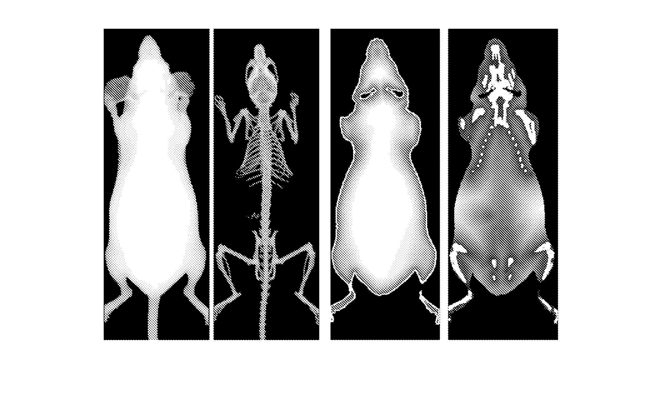
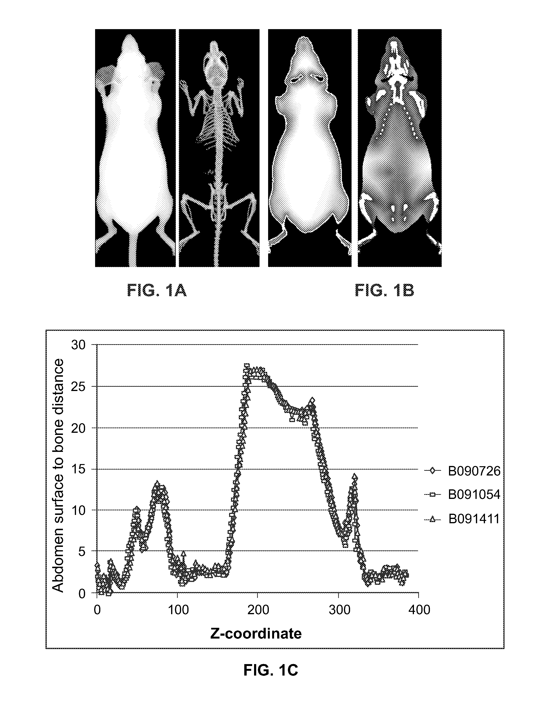
[0006]Described herein are systems and methods for efficient and accurate automated detection of a region of interest from an in vivo mammalian image. An important region of an imaged mammal is the tissue interior to the ribcage

Problems solved by technology

Acquisition of such in vivo images can be time consuming, and rapid analysis of the acquired images is key to the efficiency of the process.
The portions of an

Method used


Image

  • Method and system for automated detection of tissue interior to a mammalian ribcage from an in vivo image
  • Method and system for automated detection of tissue interior to a mammalian ribcage from an in vivo image
  • Method and system for automated detection of tissue interior to a mammalian ribcage from an in vivo image

Examples


Embodiment Construction

[0046]It is contemplated that systems, devices, methods, and processes of the claimed invention encompass variations and adaptations developed using information from the embodiments described herein. Adaptation and / or modification of the systems, devices, methods, and processes described herein may be performed by those of ordinary skill in the relevant art.

[0047]Throughout the description, where articles, devices, and systems are described as having, including, or comprising specific components, or where processes and methods are described as having, including, or comprising specific steps, it is contemplated that, additionally, there are articles, devices, and systems of the present invention that consist essentially of, or consist of, the recited components, and that there are processes and methods according to the present invention that consist essentially of, or consist of, the recited processing steps.

[0048]It should be understood that the order of steps or order for performin...


PUM


Abstract

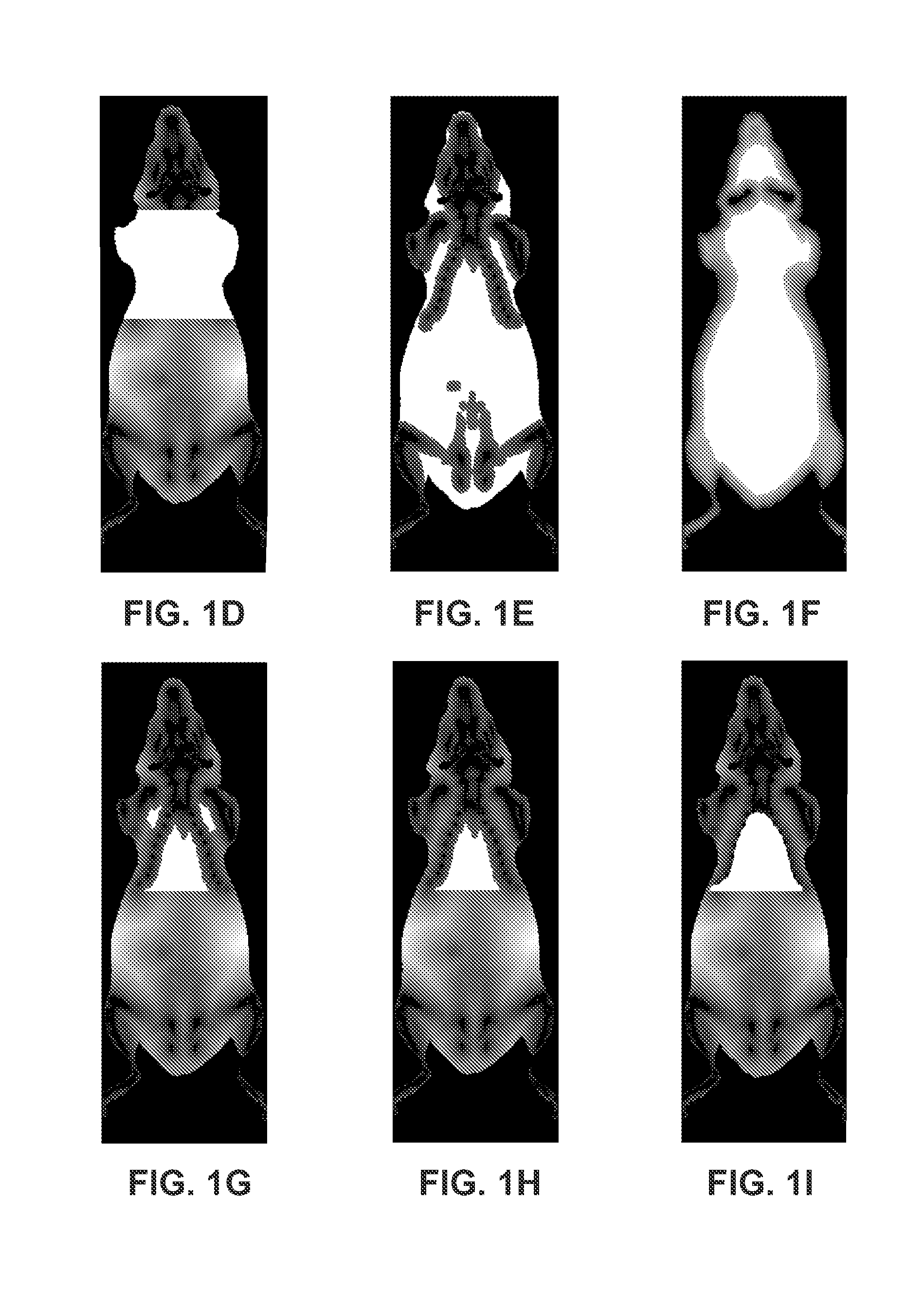
Described herein are systems and methods for efficient and accurate, automated detection of a region of interest interior to the ribcage from an in vivo mammalian image. It is found that efficient, automated identification of the region of interest interior to the ribcage can be achieved by the use of both a bone distance mask and a surface distance mask. The technique solves the problem of accurate and fast identification of the region of interest for a wide range of sizes and shapes of mammals, e.g., small mammals such as mice.

Description

TECHNICAL FIELD[0001]This invention relates generally to methods and systems of image analysis. More particularly, in certain embodiments, the invention relates to the automated detection of tissue interior to a mammalian rib cage.BACKGROUND[0002]There are a wide array of technologies directed to in vivo imaging of mammals—for example, bioluminescence, fluorescence, tomography, and multimodal imaging technologies. In vivo imaging of small mammals is performed by a large community of investigators in various fields, e.g., oncology, infectious disease, and drug discovery.[0003]In vivo imaging often involves the use of reagents, such as fluorescent probes, for non-invasive spatiotemporal visualization of biological phenomena inside a live animal. For example, fluorescence molecular tomography (FMT) involves in vivo imaging of mammals for quantitative analysis of administered and / or endogenous probes. In vivo microCT imaging, is an x-ray-based technology that can image tissues, organs, ...

Claims


Application Information

Patent Timeline
IPC IPC(8): A61B6/00
CPCA61B6/5217A61B6/508A61B6/563A61B6/505A61B6/466A61B6/469G06T2207/10072G06T2207/20152G06T2207/20156G06T2207/30008G06T7/11G06T7/155G16H50/30
Inventor OLLILAINEN, OLAVIKASK, PEET
Owner PERKINELMER CELLULAR TECH GERMANY GMBH
Why Eureka
Try Eureka