Targeting primary cilia to treat glaucoma

Active Publication Date: 2015-08-20
INDIANA UNIV RES & TECH CORP
1 Cites 0 Cited by

AI-Extracted Technical Summary

Problems solved by technology

Defective sensation of pressure may result in imbalance of aqueous humor, resulting in elevated intraocular pressure.
Low levels of eye pressure result in structural changes of the retina and poor vision, while elevated eye pressure may damage the optical nerve.
Dysfunction of the trabecular outflow leads to elevated intraocular pressure, w...

Method used

[0165]In 17-day-old wpk−/− rats, intraocular pressure was significantly reduced with systemic GS...

Benefits of technology

[0013]In one aspect, the present disclosure is directed to a method for reducing intraocular pressure ...

Abstract

Methods for reducing intraocular pressure and treating glaucoma are disclosed. The methods include administering to an individual a composition comprising a TRPV4 agonist.

Application Domain

BiocidePharmaceutical delivery mechanism +3

Technology Topic

Intraocular pressureTRPV4 +4

Image

  • Targeting primary cilia to treat glaucoma
  • Targeting primary cilia to treat glaucoma
  • Targeting primary cilia to treat glaucoma

Examples

  • Experimental program(9)

Example

Example 1
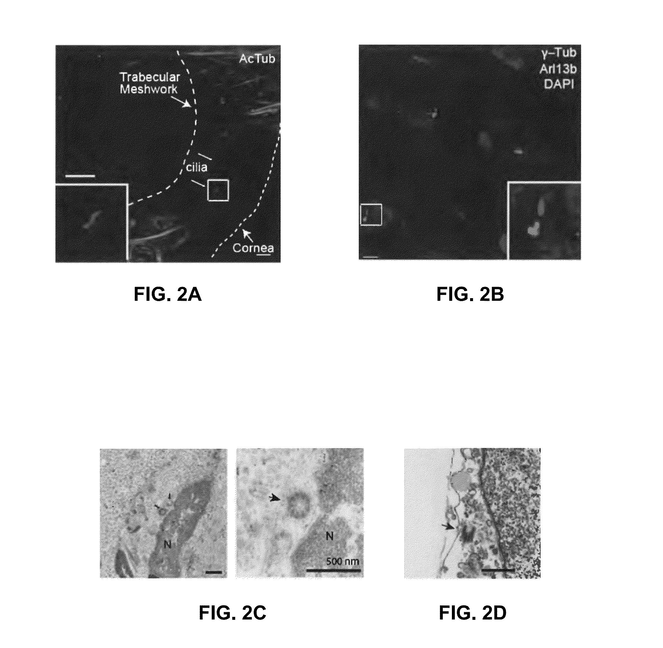
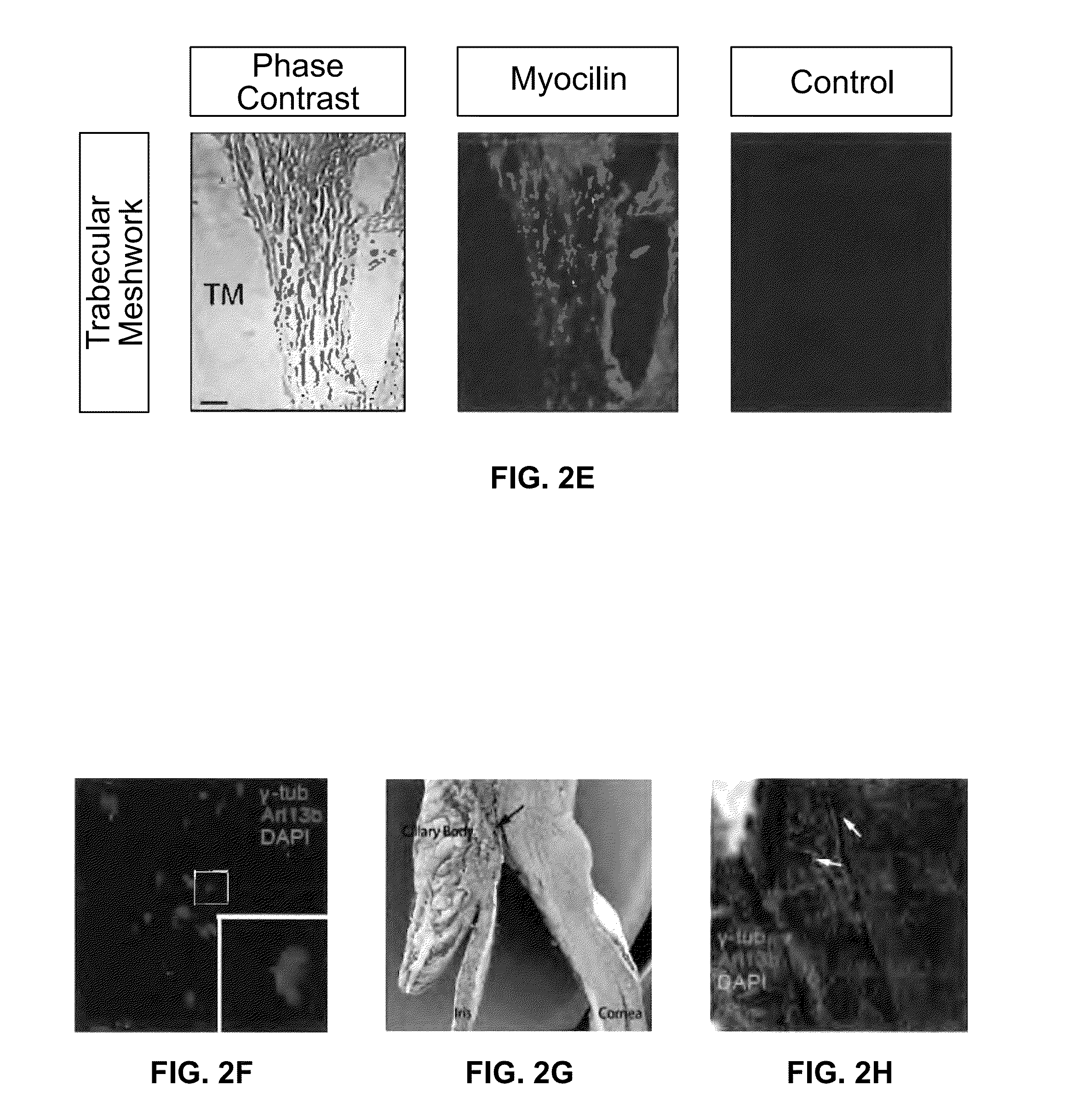
[0126]In this Example, isolated trabecular meshwork (TM) tissue from the anterior segment of human eyes was analyzed for the presence of cilia by immunofluorescence and electron microscopy.
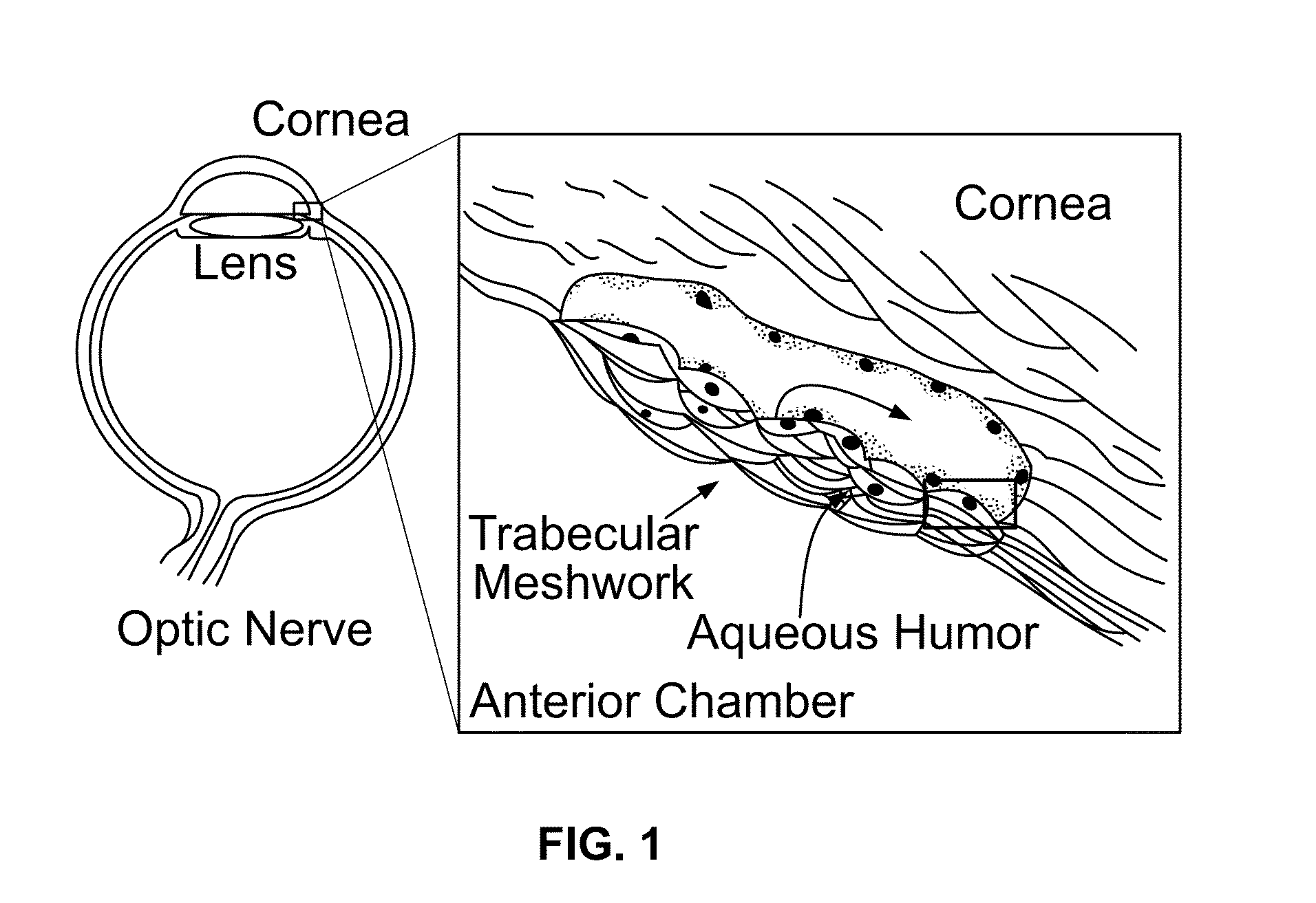
[0127]Trabecular meshwork removed from human donor eyes and surgical TM specimens during glaucoma surgery were immunostained with anti-Arl13b, acetylated α-tubulin (AcTub,), γ-tubulin (γ-Tub), and DAPI (to visualize cell nuclei). TM sections of human eyes were fixed, sectioned and electron microscopy performed. TEM images showed basal body and axonemal structures (FIG. 1, arrows).
[0128]Trabecular meshwork (TM) tissue from the anterior segment of the eye contains cilia-like structures (see schematic illustration in FIG. 1). As shown in FIGS. 2A and 2B, primary cilia were detected in TM tissues by immunostaining for acetylated α-tubulin or Arl13b, a small GTPase localized in the cilia, while the basal body was detected by staining for γ-tubulin. Collectively, this revealed numerous cilia-like structures in the uveoscleral region of the TM (FIG. 2A). The similar staining of cilia in TM tissues that were removed en face immediately following trabeculectomy from the eyes of glaucoma patients indicated that positive staining was not an artifact of fixation (FIG. 2B). The detection of primary sensory cilia with characteristic “9+0” structures by electron microscopy confirmed their presence in the TM (FIGS. 2C and 2D). Specificity for TM was verified by immunostaining of myocilin, a TM-enriched protein (FIG. 2E). Similar observations were noted in the trabecular meshwork of murine (FIG. 2F), porcine (FIGS. 2G and 2H) and bovine (FIGS. 2I and 2J) eyes. These results demonstrated that primary cilia are present in the TMs of many mammalian species.
[0129]Serum starvation is a well-described method for the induction of primary cilia. To determine whether serum starvation could induce the formation of cilia, cultured human trabecular meshwork (HTM) cells were subjected to serum starvation for 48 hours. Following serum starvation, HTM cells were immunostained to detect Arl13b, IFT88 (an intraflagellar transport protein), IFT43, IFT57, adenylate cyclase 111 (a cilia marker), and γ-tubulin (FIG. 2K).
[0130]HTM cells were then serum starved for 24 hours, 48 hours and 72 hours. As visualized in FIG. 3A, cilia length increased over time during serum starvation. Measurements of cilia demonstrated that the average lengths of cilia increased following serum deprivation. At 24 hours the cilia lengths were 2.2±0.7 μm; at 48 hours, 4.3±0.6 μm; and were 5.6±0.4 m at 72 hours (FIG. 3B). As illustrated in FIG. 3C, ciliation rate also increased over time. These results strongly confirmed the presence of functional primary cilia in trabecular meshwork cells.

Example

Example 2
[0131]In this Example, the effect of pressure changes on cilia length in TM cells was determined.
[0132]HTM cells were serum starved for 48 hours, cultured in a pressure chamber at 0 mmHg, 30 mmHg and 50 mmHg pressure for 0 hour, 1 hour and 3 hours. After cell fixation, cilia were imaged by immunostaining for acetylated α-tubulin. As shown in FIG. 4A, cilia length was reduced by pressure. Average cilia lengths were 5.3±0.2 μm in the normal pressure groups and 3.9±0.2 μm in the high pressure (30 mmHg and 50 mmHg) groups (FIG. 4B). Ciliary length reduction by pressure occurred in a time-dependent fashion (FIG. 4C). Changes in ciliary length were both time- and dose-dependent processes.
[0133]TNFα and TGFβ are both highly abundant in the aqueous humor of patients with acute and chronic open angle glaucoma. To determine whether the expression of TNFα was upregulated in response to pressure, its transcript levels were measured by qRT-PCR under different pressures.
[0134]HTM cells were serum-starved for 48 hours and treated with saline, saline plus 50 mmHg pressure, chlorohydrate, and chlorohydrate plus 50 mmHg pressure for 3 hours. Levels of mRNA of TNFα, TGFβ and Gli1 were then measured. Results presented represent three independent experiments. Serum-starved HTM cells were also placed under 50 mmHg pressure for 0, 10, 20, 40, 60 and 80 minutes and analyzed for expression of TNFα and IL-33. Results presented represent three independent experiments.
[0135]Following 60 minutes of application of 50 mmHg pressure, which mimics acute angle closure glaucoma, a robust elevation of TNFα transcription was observed (FIG. 4D). The lack of changes in IL-33 transcript indicated that the effects on TNFα are specific (FIG. 4E). Further, application of 50 mmHg pressure markedly elevated the transcript levels of TNFα and TGFβ1, but had modest effects on Gli1, a factor in the Sonic Hedgehog pathway that requires primary cilia for signaling (FIG. 4D). The dependence of these effects on functional cilia was then determined by testing their sensitivity to chlorohydrate, which effectively removes cilia. Treatment of cells with chlorohydrate reduced the elevation of gene transcription in response to pressure (FIG. 4D).
[0136]In a complementary approach, cilia were ablated by shRNA knockdown of Rab8, which inhibits ciliogenesis. Cells with reduced expression of Rab8 also showed a reduction of gene transcription (Rab8 and OCRL) in response to pressure (FIGS. 4F-4H).
[0137]Together, these experiments strongly suggested that enhanced transcription of TNFα and TGFβ1 by pressure occurs via a cilia-dependent process.

Example

Example 3
[0138]To further characterize the distribution of ciliary proteins under elevated pressure conditions, intraflagellar transport (IFT) proteins were examined.
[0139]The distribution of proteins involved in both anterograde (IFT57, IFT88) and retrograde (IFT20, IFT43, IFT144) ciliary trafficking were examined using immunohistology. Specifically, HTM cells were serum-starved to induce ciliogenesis, followed by treatment with or without elevated pressure (50 mmHg). Representative images of immunostaining with antibodies for IFT88 (green), acetylated α-tubulin (red), and DAPI (blue) show the loss of IFT88 in the distal tip of cilia under elevated pressure conditions (FIGS. 5 and 6). Only IFT88 was found to have a markedly altered distribution under increased pressure conditions. In control HTM cells, IFT88 distributed to the base as well as to the tip of the axoneme; in HTM cells treated with pressure, IFT88 accumulated at the base of the cilia with a marked decrease at the ciliary tip.
[0140]Thus, elevated pressure in cells alter IFT distribution with a resultant change in ciliary protein trafficking.

PUM

PropertyMeasurementUnit
Dimensionless property5.0E-8dimensionless
Dimensionless property5.0E-7dimensionless
Pressure

Description & Claims & Application Information

We can also present the details of the Description, Claims and Application information to help users get a comprehensive understanding of the technical details of the patent, such as background art, summary of invention, brief description of drawings, description of embodiments, and other original content. On the other hand, users can also determine the specific scope of protection of the technology through the list of claims; as well as understand the changes in the life cycle of the technology with the presentation of the patent timeline. Login to view more.

Similar technology patents

Intraocular physiological sensor

PendingUS20200305741A1improve usability and overall lifespanreduce intraocular pressure
Owner:GLAUKOS CORP

Classification and recommendation of technical efficacy words

  • reduce intraocular pressure

Intraocular physiological sensor

PendingUS20200305741A1improve usability and overall lifespanreduce intraocular pressure
Owner:GLAUKOS CORP
Why Eureka
Try Eureka