Surgical assistance system

a technology of assistance system and surgical procedure, applied in the field of surgical assistance system, can solve the problems of poor quality of sections obtained from frozen tissue samples, high demands on the ability of pathologists, and good pathologists to return erroneous analyses, etc., and achieve the effect of reducing the time required for preparing the pathological diagnosis and expanding the possibility of cooperation

Active Publication Date: 2015-09-10
CARL ZEISS MEDITEC AG
View PDF6 Cites 11 Cited by
  • Summary
  • Abstract
  • Description
  • Claims
  • Application Information

AI Technical Summary

Benefits of technology

[0023]In this case, the functional control data contain control data for controlling the image capture device having a hyperspectral sensor. Such a sensor allows the pathologist to conduct a thorough spectral analysis of the light reflected by the tissue to be examined.
[0024]When the imaging device comprises one or more of the functional units listed above, the diagnostic information available to the pathologist is considerably expanded. In particular, when a plurality of the functional units listed above are present, the pathologist has the ability to obtain a comprehensive view of the tissue to be examined. This can considerably reduce the risk of a misdiagnosis.
[0028]Within the framework of the invention, the surgical assistance system may also be designed in such a way that it is able to provide an automated diagnosis. In such a case, the pathology unit comprises a diagnostic database that contains a number of stored diagnoses as well as a number of stored image data and in which each of the number of stored diagnoses is associated with at least one characteristic piece of image data out of the number of stored image data. In addition, the pathology unit then comprises an image analysis unit for the extraction of particular image data from the cellular-level image data and / or overview image data received as well as a diagnostic unit that is connected to the image analysis unit for the purpose of receiving the extracted image data and connected to the diagnostic database for the purpose of receiving the stored image data. On the basis of a comparison of the extracted image data to the received stored image data, the diagnostic unit selects a diagnosis and displays it on the display unit as a suggested diagnosis. This allows a preliminary recommended diagnosis to be quickly provided to the pathologist. If the comparison shows that certain image data match the characteristic image data for multiple diagnoses, a preliminary suggestion of possible diagnoses may be offered to the pathologist. The pathologist is then able in a first step to review the diagnoses offered and either verify or discard them. By pre-selecting possible diagnoses, the time required for preparing the pathological diagnosis may be shortened.
[0029]The surgical assistance system according to the invention offers the pathologist a full range of options for influencing the gathering of data and / or providing information to the treating surgeon. As compared to the diagnostic systems known from the prior art, the surgical assistance system according to the invention expands options for cooperation between the pathologist and the treating surgeon.

Problems solved by technology

Because the quality of sections acquired from frozen tissue samples is poorer than the quality of sections of formalin-fixed, paraffin-embedded tissues and because only a limited amount of time is available for analysis, the demands on the abilities of the pathologist are very high.
However, by the nature of the system, due to the low number of tissue samples, the poor quality of the frozen sections, and the short period of time available for analysis, it is possible for even a very good pathologist to return erroneous analyses relatively frequently, even when experienced pathologists are able to attain accuracy rates of 90% or more.

Method used


Image

  • Surgical assistance system
  • Surgical assistance system
  • Surgical assistance system

Examples


Embodiment Construction

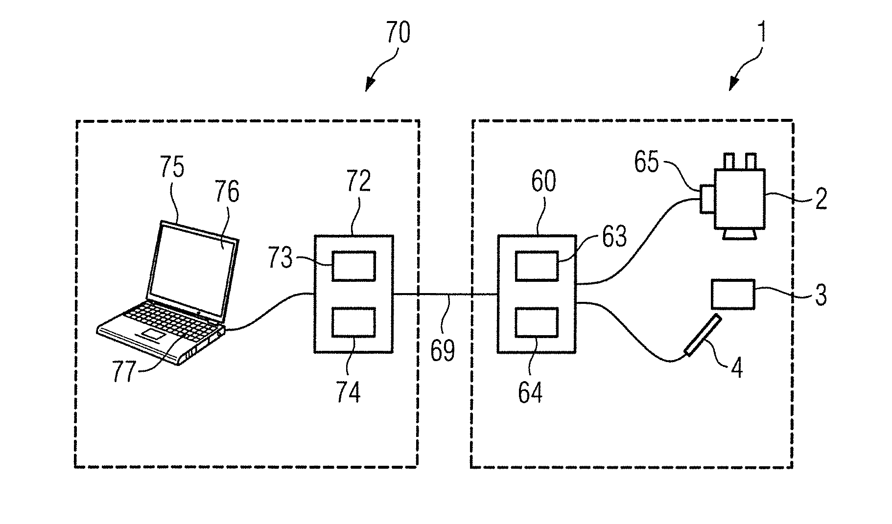
[0038]A first exemplary embodiment of the surgical assistance system according to the invention will be described below with reference to FIG. 1. FIG. 1 schematically depicts a group of devices 1 located in the operating room as well as a pathology unit 70 that may be located outside the operating room. Typically, the pathology unit 70 is located at a distance from the operating room in a different room of the same building. However, it is also fundamentally possible for the pathology unit to be located in a different building or even in a different city or another country.

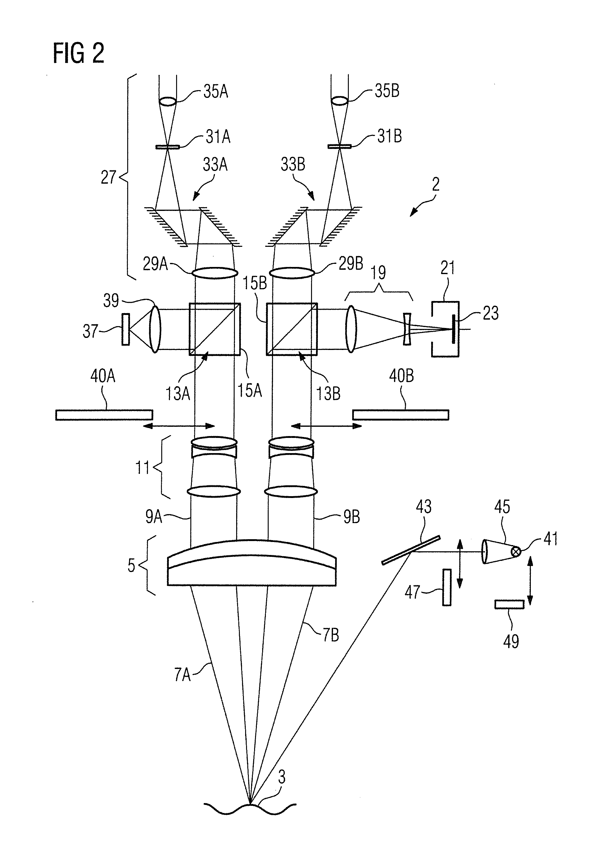
[0039]The group of devices 1 comprises a surgical microscope 2 by means of which an operating field 3 may be observed. Using the surgical microscope 2, essentially an overview image of the operating field 3 is acquired. Moreover, the group of devices 1 comprises an endomicroscope 4 by means of which cellular-level image data may be captured at selected locations in the operating field 3. The surgical microscope 2 ...


PUM


Abstract

A surgical assistance system includes an endomicroscope (4) and an imaging device (2) for capturing image data, and a transmission unit (63) for transmitting the image data. A pathology unit (70) includes a receiving unit (74) for receiving the image data and a display unit (76) displays the image data. A data connection (69) connects the transmission unit (63) to the receiving unit (74). The imaging device (2) and / or the endomicroscope (4) has at least one functional unit assigned to a functional control unit (65). A data receiving unit (64) receives data from the pathology unit (70). The pathology unit (70) has an input unit (77) for inputting functional control data for the functional control unit (65) and a transmitter unit (73) transmits the functional control data to the devices. The data receiving unit (64) receives and relays the functional control data to the functional control unit (65).

Description

BACKGROUND[0001]1. Field of the Invention[0002]The present invention relates to a surgical assistance system and, in particular, to an assistance system for microsurgery.[0003]2. Description of the Related Art[0004]In surgical interventions and, in particular, in microsurgical interventions and tumor operations, intraoperative tissue samples are taken, which are then analyzed by a pathologist using frozen section diagnosis. The results of this analysis are then relayed to the operating surgeon by telephone and analyzed by the surgeon for the purpose of determining the further course of treatment. In order to perform such a frozen section diagnosis, the tissue samples acquired intraoperatively are sense to a pathology laboratory, where the samples are analyzed. There, the tissue samples are frozen and sectioned before being analyzed by the pathologist. This process generally takes around 15 minutes for one tissue sample. The period of time until the results of the analysis are receiv...

Claims


Application Information

Patent Timeline
Patent Type & Authority Applications(United States)
IPC IPC(8): A61B5/00A61B17/00A61B1/313A61B19/00A61B1/00G16H20/40G16H30/20G16H40/67G16H50/20
CPCA61B5/0084A61B5/0071A61B19/5223A61B5/0075A61B19/5244A61B2017/00973A61B1/313A61B17/00234A61B5/0035A61B2019/5257A61B2019/5265A61B1/00009A61B90/25A61B90/20A61B2034/2065G16H40/67G16H20/40G16H30/20G16H50/20A61B1/000094
Inventor HAUGER, CHRISTOPHGUCKLER, ROLANDPANITZ, GERALD
Owner CARL ZEISS MEDITEC AG
Why Eureka
Try Eureka