Method for building a 3D model of a rock sample

a 3d model and rock technology, applied in the direction of material analysis using wave/particle radiation, image enhancement, instruments, etc., can solve the problems of thin sections as sources, destructive sections at microscale, and the boundary between dark and bright regions is not step-lik

Active Publication Date: 2015-09-17
SCHLUMBERGER TECH CORP
View PDF4 Cites 23 Cited by
  • Summary
  • Abstract
  • Description
  • Claims
  • Application Information

AI Technical Summary

Benefits of technology

[0008]A method for building a 3D model of a rock sample comprises performing X-ray micro / nanoCT scanning of a rock sample and obtaining its initial three-dimensional microstructure image in a gray scale. Then, an analysis of the obtained three-dimensional image of the rock sample is performed and a binarization (multicomponent segmentation) method is selected in dependence of the image quality and properties of the sample. The selected binarization method is at least once applied to the obtained initial three-dimensional image of the sample. Obtained 3D binarized image represents a 3D model of the rock sample.

Problems solved by technology

The problem is that the boundary between dark and bright regions is not step-like.
Main disadvantage of this application is presence of 2D-3D image registration as necessary step for building the 3D digital model accounting pore space and mineral distribution.
Another disadvantage is necessity of making thin sections as sources for 2D mineral maps from SEM or optical microscopy.
It is known that making this sections is destructive at microscale.
This makes their 2D-3D registration procedure nearly non-applicable in real life.
Furthermore, preparing thin sections from the sample after X-Ray microCT destructs original sample, so no further investigations with original sample are possible.

Method used


Image

  • Method for building a 3D model of a rock sample
  • Method for building a 3D model of a rock sample
  • Method for building a 3D model of a rock sample

Examples


Embodiment Construction

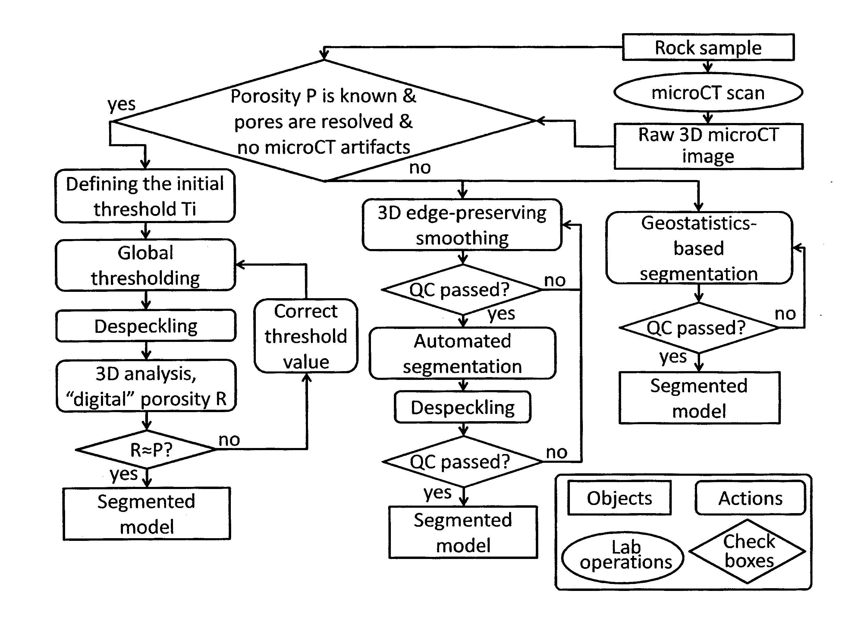
[0015]Detailed flowchart illustrating an exemplary 3D model building method is shown on FIG. 4. In the present example, rock samples (core plugs, drilling cuttings, other rocks) are transported to a computer tomographic (“CT”) scanner, which may use x-rays for analysis of internal structure of the samples and for generation of three dimensional (3D) images of the samples. An X-ray micro / nanoCT scanning of a rock sample is performed at a particular resolution and a 3D initial image in gray scale is obtained.

[0016]Then analysis of the obtained initial three-dimensional image of the rock sample is performed, namely the presence of significant artifacts (microCT ring artifacts, smoothing due to thermal drifts of an X-ray source, beam hardening artifact, partial volume effects, signal-to-noise levels) is checked. A binarization method is selected in dependence of the image quality and properties of the rock sample. Image binarization refers to the process of converting an image represent...


PUM


Abstract

A method for building a 3D model of a rock sample comprises performing X-ray micro/nanoCT scanning of a rock sample and obtaining its initial three-dimensional microstructure image in a gray scale. Then, an analysis of the obtained three-dimensional image of the rock sample is performed and a binarization method is selected in dependence of the image quality and properties of the rock sample. The selected binarization method is at least once applied to the obtained initial three-dimensional image of the sample. Obtained 3D binarized image represents a 3D model of the rock sample.

Description

FIELD OF THE INVENTION[0001]The invention relates to X-ray based analysis of a rock sample, namely microtomography (microCT) and nanotomography (nanoCT) techniques.BACKGROUND OF THE INVENTION[0002]X-ray micro- and nano- computed tomography is a well-known non-destructive technique for visualizing and quantifying the internal structure of objects in three dimensions (3D). It is used to provide high resolution images of rocks in 2D or 3D at a micron scale (see, for example, M. A. Knackstedt et al. , “Digital Core Laboratory: Properties of Reservoir Core Derived From 3D Images,” SPE 87009, 2004).[0003]X-ray micro- and nano- computed tomography techniques enable acquisition of grayscale 3D images in which the grayscale represents the X-ray absorption distribution within the object. The absorption (attenuation) depends on the chemical composition of the material and its physical density. The range of X-ray energies used in computed tomography (CT) allows the study of very dense objects, ...

Claims


Application Information

Patent Timeline
Patent Type & Authority Applications(United States)
IPC IPC(8): G06T17/05G06T5/20G06T7/00
CPCG06T17/05G06T7/00G06T5/20G06T2207/20032G06T2207/10116G06T2207/30184G06T2207/10081G01N23/046G01N33/24G01N2223/419G01N2223/616G01N2223/649G06T5/002G06T5/40G06T7/11G06T2207/20192
Inventor KOROTEEV, DMITRY ANATOLIEVICHNADEEV, ALEXANDER NIKOLAEVICHYAKIMCHUK, IVAN VIKTOROVICHVARFOLOMEEV, IGOR ANDREEVICH
Owner SCHLUMBERGER TECH CORP
Why Eureka
Try Eureka