MR Imaging Apparatus And Method For Generating A Perfusion Image With Motion Correction

a technology of perfusion image and imaging apparatus, which is applied in the field of method and system for generating magnetic resonance images, can solve the problems of high-resolution 3d mr imaging being susceptible to motion corruption, mr images based on asl differences often suffer from image noise, and can be noisy and noisy,

Active Publication Date: 2015-10-01
SIEMENS HEALTHCARE GMBH +1
View PDF4 Cites 27 Cited by
  • Summary
  • Abstract
  • Description
  • Claims
  • Application Information

AI Technical Summary

Benefits of technology

[0011]Exemplary embodiments of the present disclosure can provide a magnetic resonance system for generating real-time (prospective) motion-corrected perfusion images based on pulsed arterial spin labeling (PASL). The MR system can provide prospective motion correction, for e

Problems solved by technology

The difference in the MR signal intensity for labeled and control images is typically only a few percent of the tissue MR signal, and thus MR images based on ASL differences often suffer from the influence of image noise.
Such factors of imaging time and small signals can make ASL techniques prone to motion corruption and artifacts, which can a

Method used


Image

  • MR Imaging Apparatus And Method For Generating A Perfusion Image With Motion Correction
  • MR Imaging Apparatus And Method For Generating A Perfusion Image With Motion Correction
  • MR Imaging Apparatus And Method For Generating A Perfusion Image With Motion Correction

Examples


example

[0062]The system and method described herein were tested in a healthy human volunteer to assess the efficacy of the disclosed 3D navigator-based real-time motion correction for artery spin labeled imaging using a 3D GRASE readout. The imaging of blood perfusion activity in the subject's brain was done using a 3T MR scanner (MAGNETOM Skyra, Siemens Healthcare, Erlangen) with a 32 channel head coil.

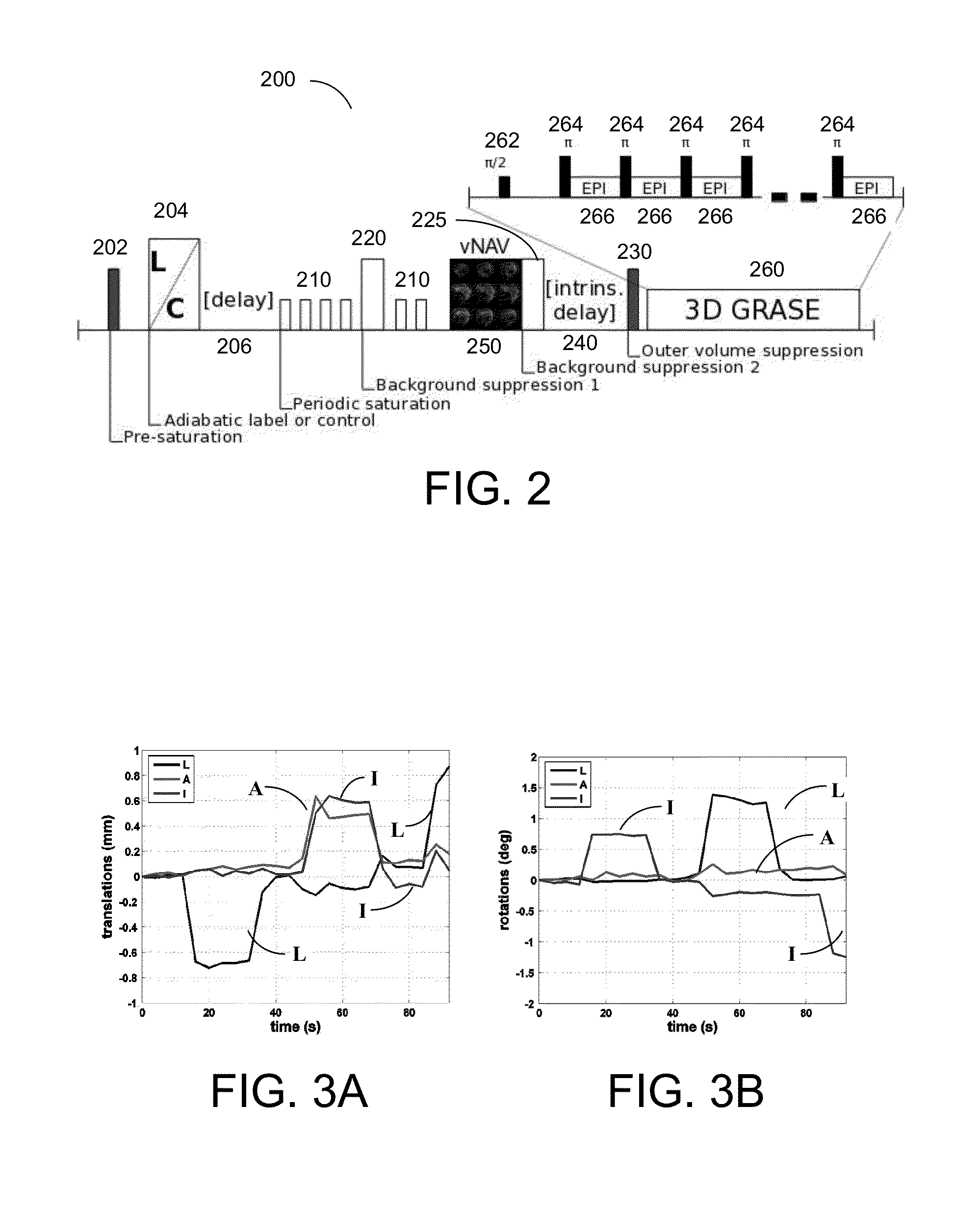
[0063]A pulse sequence substantially corresponding to that illustrated in FIG. 2 was used for generating and acquiring perfusion image data. This sequence 200 includes a 3D segmented GRASE readout sequence 260 with pulsed ASL (PASL) preparation that includes flow-sensitive alternating inversion recovery with a quantitative imaging of perfusion using a single subtraction, second version, with thin-slice T1 periodic saturation (FAIR Q2TIPS), as described in the Luh et al. reference cited herein. Exemplary parameters that were used for the imaging procedure were: TR=4 s; TI=2.4 s (for gray / whi...


PUM


Abstract

A magnetic resonance method and system are provided for generating real-time motion-corrected perfusion images based on pulsed arterial spin labeling (PASL) with a readout sequence such as a 3D gradient and spin echo (GRASE) image data acquisition block. The real-time motion correction is achieved by using a volumetric 3D EPI navigator that is provided during an intrinsic delay in the PASL sequence, which corrects for motion prospectively and does not extend the image data acquisition time as compared to a similar non-motion-corrected imaging procedure.

Description

CROSS-REFERENCE TO RELATED APPLICATION(S)[0001]The present application relates to and claims priority from U.S. Provisional Patent Application Ser. No. 61 / 973,324 filed Apr. 1, 2014, the disclosure of which is incorporated herein by reference in its entirety.STATEMENT REGARDING FEDERALLY SPONSORED RESEARCH[0002]The present invention was made with government support under Grants R21MH096559, R01HD071664, R21EB008547, R33DA026104, and P41RR014075 awarded by the National Institute of Health (NIH). The government has certain rights in the invention.FIELD OF THE DISCLOSURE[0003]The present disclosure relates to a method and a system for generating magnetic resonance images, and in particular to a method and a system for generating magnetic resonance 3D perfusion images with motion correction.BACKGROUND INFORMATION[0004]Magnetic resonance (MR) imaging is a known technology that can produce images of the inside of an examination subject without radiation exposure. In a typical MR imaging p...

Claims


Application Information

Patent Timeline
IPC IPC(8): A61B5/026G01R33/563G01R33/561A61B5/055
CPCA61B5/0263A61B5/055G01R33/5618G01R33/5635G01R33/56366A61B5/7207G01R33/5616G01R33/5676
Inventor HEBERLEIN, KEITH AARONBHAT, HIMANSHUTISDELL, MATTHEW DYLANVAN DER KOUWE, ANDRE JAN WILLEM
Owner SIEMENS HEALTHCARE GMBH
Why Eureka
Try Eureka