Automated imaging of chromophore labeled samples

Active Publication Date: 2015-11-12
CELLOMICS
View PDF8 Cites 22 Cited by
  • Summary
  • Abstract
  • Description
  • Claims
  • Application Information

AI Technical Summary

Benefits of technology

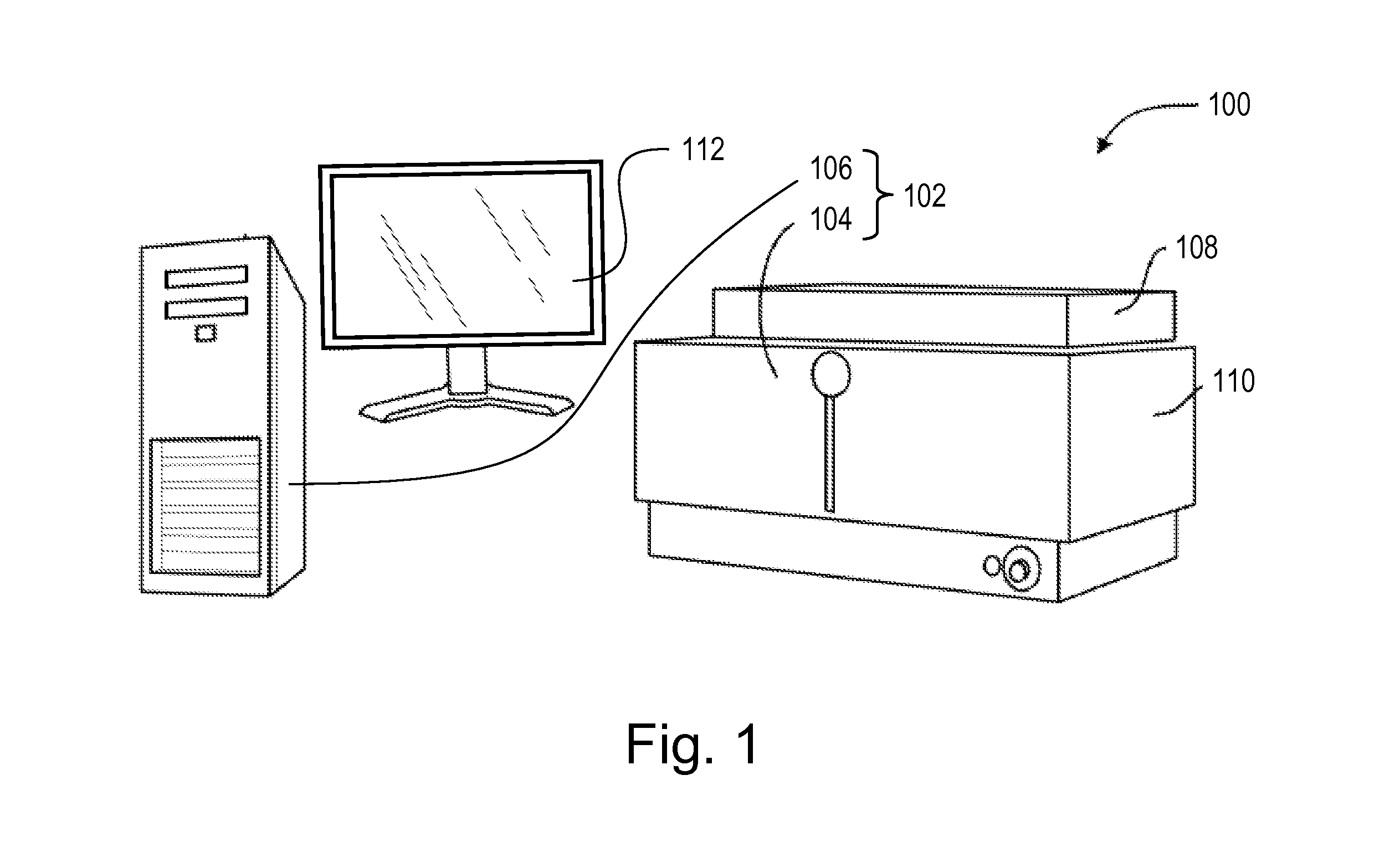
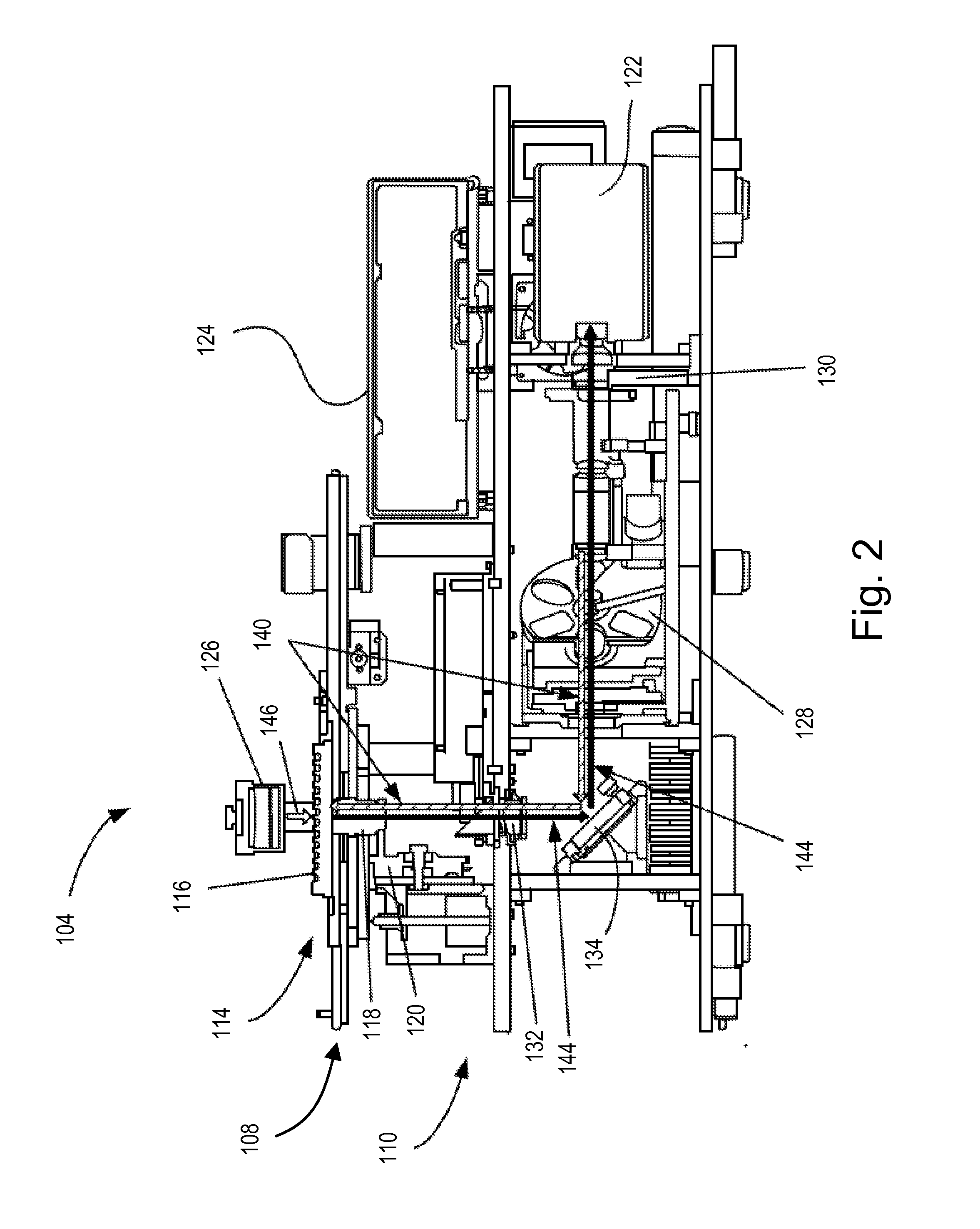
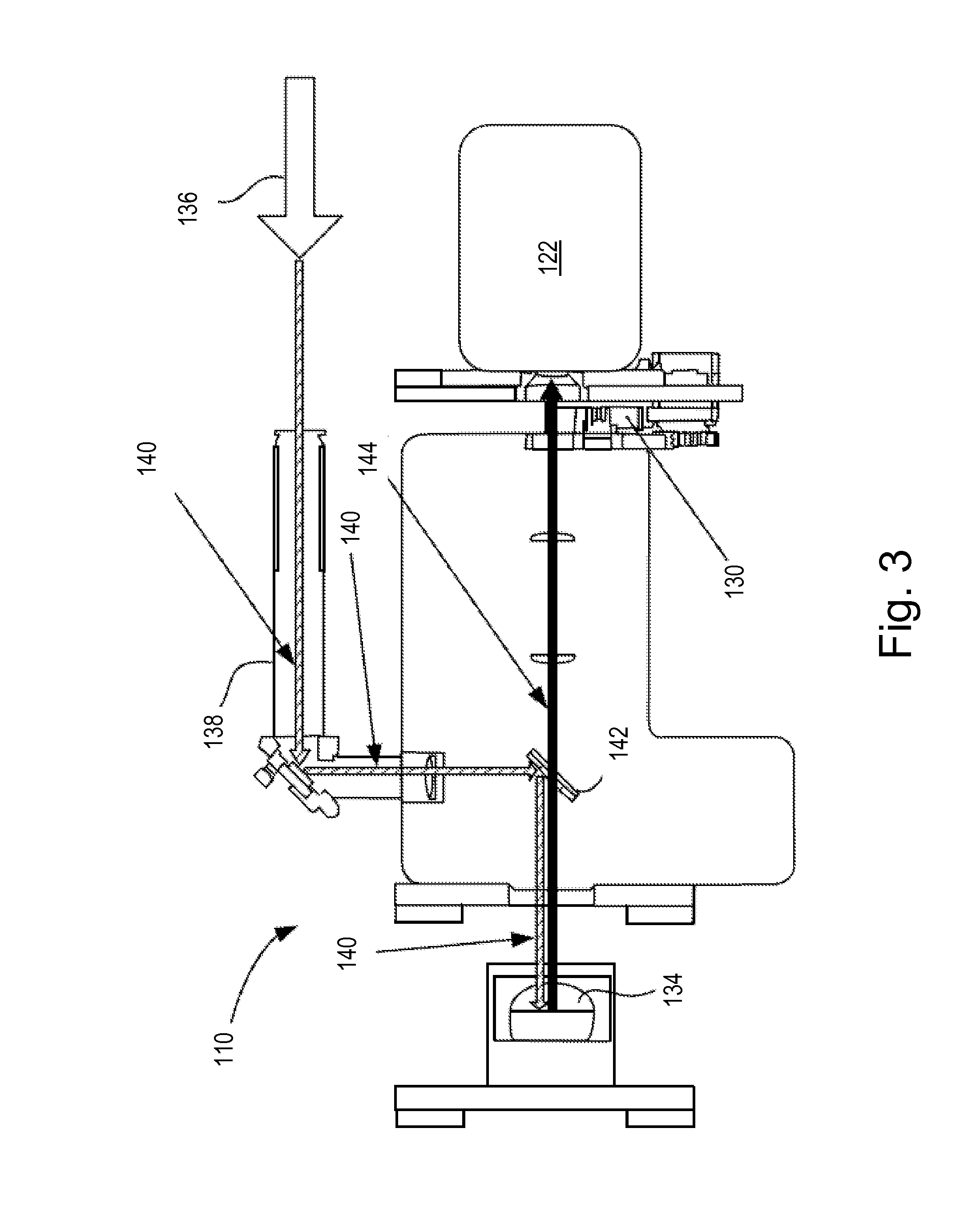
[0016]Various embodiments disclosed herein are related to a High-Content Imaging (HCl) System that provides for HCl on cell and/or tissue samples labeled with fluorescence probes that can also be labeled with chromophores. Furthermore, the ability of HCl to multiplex different imaging modes means that any combination of three modes of operation, as detail

Problems solved by technology

However, the drawback to using brightfield microscopy when applied to biological samples is its poor contrast.
It is to be also noted that automated tissue scanners for digital pathology have the ability to image both fluorescence and chromophore absorbance bu

Method used


Image

  • Automated imaging of chromophore labeled samples
  • Automated imaging of chromophore labeled samples
  • Automated imaging of chromophore labeled samples

Examples


Embodiment Construction

[0035]As used in the specification, a word appearing in the singular encompasses its plural counterpart, and a word appearing in the plural encompasses its singular counterpart, unless implicitly or explicitly understood or stated otherwise. Furthermore, it is understood that for any given component or embodiment described herein, any of the possible candidates or alternatives listed for that component may generally be used individually or in combination with one another, unless implicitly or explicitly understood or stated otherwise. Additionally, it will be understood that any list of such candidates or alternatives is merely illustrative, not limiting, unless implicitly or explicitly understood or stated otherwise. In addition, unless otherwise indicated, numbers expressing quantities of ingredients, constituents, reaction conditions and so forth used in the specification and claims are to be understood as being modified by the term “about.”

[0036]Accordingly, unless indicated to ...


PUM


Abstract

A system and method that images biological samples and uses chromophores to analyze the imaged samples. The chromophore analysis can be done by itself or in conjunction with fluorophore analysis in High Content Imaging systems. To perform chromophore analysis the biological samples can be labeled with different chromophores and imaged using transmitted light that is at least partially absorbed by the chromophores. To also perform fluorophore analysis the samples can also be labeled with fluorophores that are excited by excitation light. The chromophore analysis and fluorophore analysis can be performed separately or concurrently using a High Content Imaging system. The system provides the expanded capability by illuminating the chromophore-labeled samples with transmitted light of different wavelengths and automatically detecting the images which represent the differential absorption of the colored lights by the sample.

Description

CROSS REFERENCE TO RELATED APPLICATIONS[0001]This application claims priority to U.S. Provisional Patent Application No. 61 / 992,008, filed on May 12, 2014, the contents of which are hereby incorporated by reference.BACKGROUND[0002]1. Field[0003]The present application relates to the use of automated imaging systems for high-content screening and analysis, collectively referred to as high-content imaging. More particularly, the present application relates to high-content imaging using a system having multiplexed imaging capabilities that include imaging of different chromophore labeled samples.[0004]2. the Relevant Technology[0005]A growing trend in microscopy over the last decade is the automated imaging of biological samples. Rather than the manual observation of samples, automated microscopy involves computer-controlled automatic selection and digital imaging of sample fields, enabling high throughput imaging of a large number of samples without end-user input.[0006]Automated imag...

Claims


Application Information

Patent Timeline
IPC IPC(8): G01N21/64G01N33/487G02B5/02
CPCG01N21/6486G02B5/0278F21Y2105/003G01N33/487G02B5/0294G01N21/253G01N21/6452G01N21/6458G01N21/255G01N2201/0627G01N2021/6491F21Y2105/10G01N2201/0631G01N2201/064F21Y2105/14G01N21/31G01N21/6428G01N35/00029G01N2021/6439G01N2035/00158G02B21/16G02B21/367
Inventor GHOSH, RICHIK NILOYVANDENBERG, III, DIRK JOHNHEFFLEY, KEITH RAOTOMASZEWSKI, MONICA JOHASKINS, JEFFREY ROBERT
Owner CELLOMICS
Why Eureka
Try Eureka