Method and device for ultrasound guided minimal invasive access of a bodily cavity

a minimal invasive and ultrasound guided technology, applied in the field of ultrasound guided minimal invasive access of the bodily cavity, can solve the problems of major morbidity and mortality, inadvertent bowel or vascular injury, and injury of the aforementioned structures, and achieve the effect of preventing entry injuries and better ultrasound views

Inactive Publication Date: 2015-11-19
ESANU CATALIN
View PDF9 Cites 6 Cited by
  • Summary
  • Abstract
  • Description
  • Claims
  • Application Information

AI Technical Summary

Benefits of technology

[0014]In accordance with the present invention, a puncture assistance device for use in connection with an ultrasound probe to guide a Veress needle through the layers of the wall into a bodily cavity is provided. The puncture assistance device comprises a tenaculum-like forceps provided with ratchet fixation and sharp incurving, needle like pointed blades, a needle guide body provided with a slot for guiding the Veress needle within the scan plane of the ultrasound probe, attachment means to the ultrasound probe, and attachment means of the needle guide to the tenaculum-like forceps. The improvement consists in the possibility of maneuvering the cavity wall layers by the tenaculum-like forceps, into spacing them apart for better ultrasound view and to create access space for preventing entry injuries.
[0015]Pursuant to another feature of the present invention, a balloon catheter for use as a retractor in gaining access into the abdominal cavity, is comprising an elongated shaft with a balloon retractor at the distal end of the elongated shaft, providing an inner balloon membrane that is inflatable to a maximum volume and an attached outer framework of non-distendable flexible fibers disposed in a specific arrangement on the different faces of the balloon, providing a mesh-like structure on a side of the balloon. A pilot balloon is connected to the balloon retractor. The balloon retractor is inserted guided by the Veress needle through the layers of the abdomen with balloon in a collapsed state, followed by its inflation, leading to the expansion of the fiber net, and upon piercing by a sharp trocar through the layers of the abdomen and balloon, the burst of said balloon is followed by the entanglement of the tip of the trocar in the mesh, preventing thereby puncturing wounds to the internal organs made by the sharp tip of the trocar upon insertion.
[0016]Another aspect of the current invention is represented by a method to develop pneumoperitoneum with the aid of an ultrasound device. This is comprising making an incision in the skin of the abdominal wall, attaching the puncturing assistance device that is operatively connected to an ultrasound transducer to the abdominal fascia, inserting a Veress needle through the said puncturing device, observing the path of the Veress needle through the layers of the a

Problems solved by technology

This may result in inadvertent bowel or vascular injury which can be responsible for major morbidity and mortality.
The problem resides in the proximity of the large retroperitoneal vessels and the frequency of infraumbilical adhesions by the bowel and omentum found in almost 10% of cases, which can lead to injury of the aforementioned structures, especially in the case of a previous laparotomy or obese or too thin patients.
Although it takes less time to perform than the Veress needle technique, and is associated with less mino

Method used


Image

  • Method and device for ultrasound guided minimal invasive access of a bodily cavity
  • Method and device for ultrasound guided minimal invasive access of a bodily cavity
  • Method and device for ultrasound guided minimal invasive access of a bodily cavity

Examples


Embodiment Construction

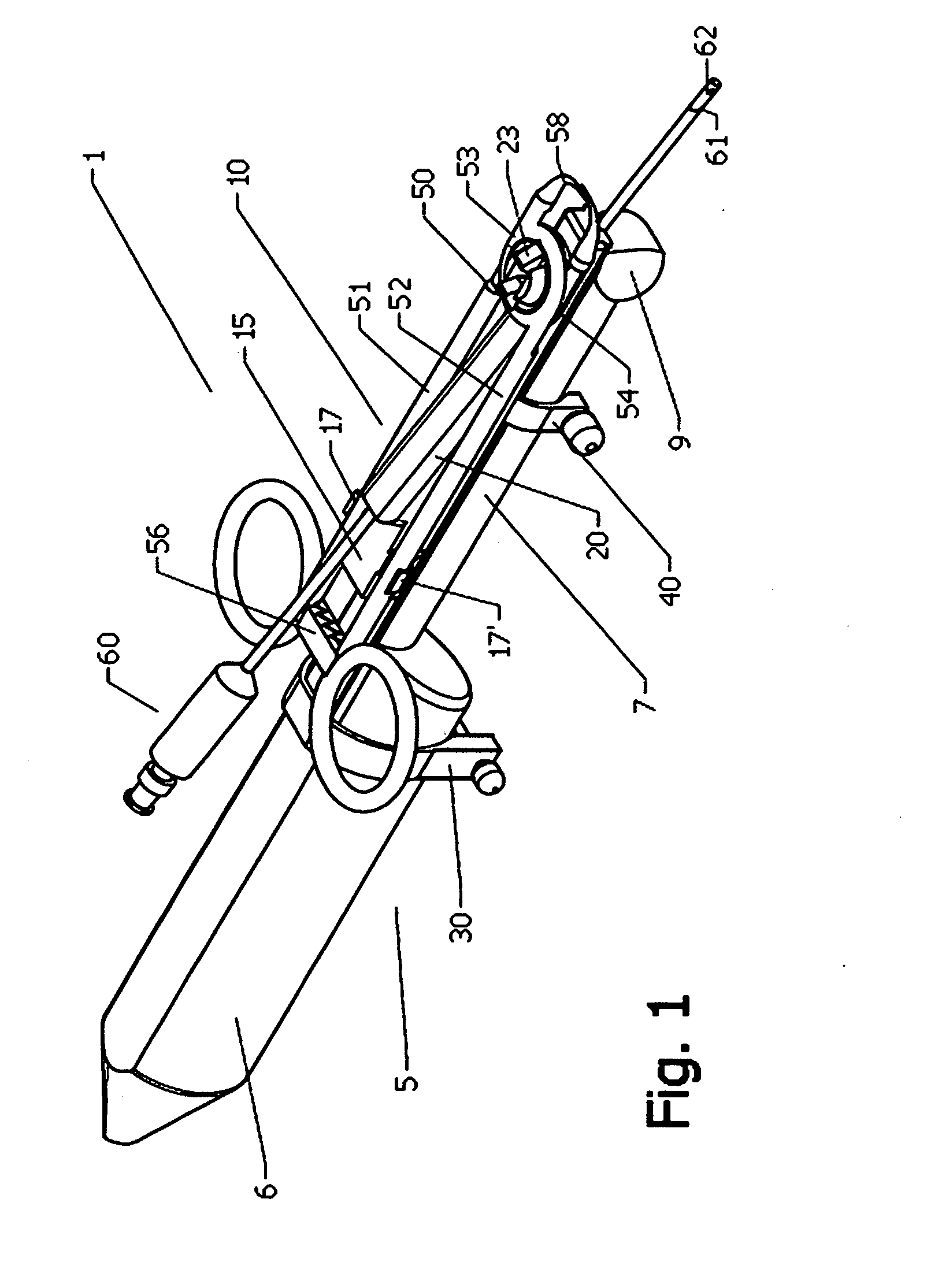
[0039]In the following specification I shall nominate as proximal a part of the assembly that is located relatively close to the operator, and as distal a part of the assembly that is located further away from the operator and hence close to the operating field.

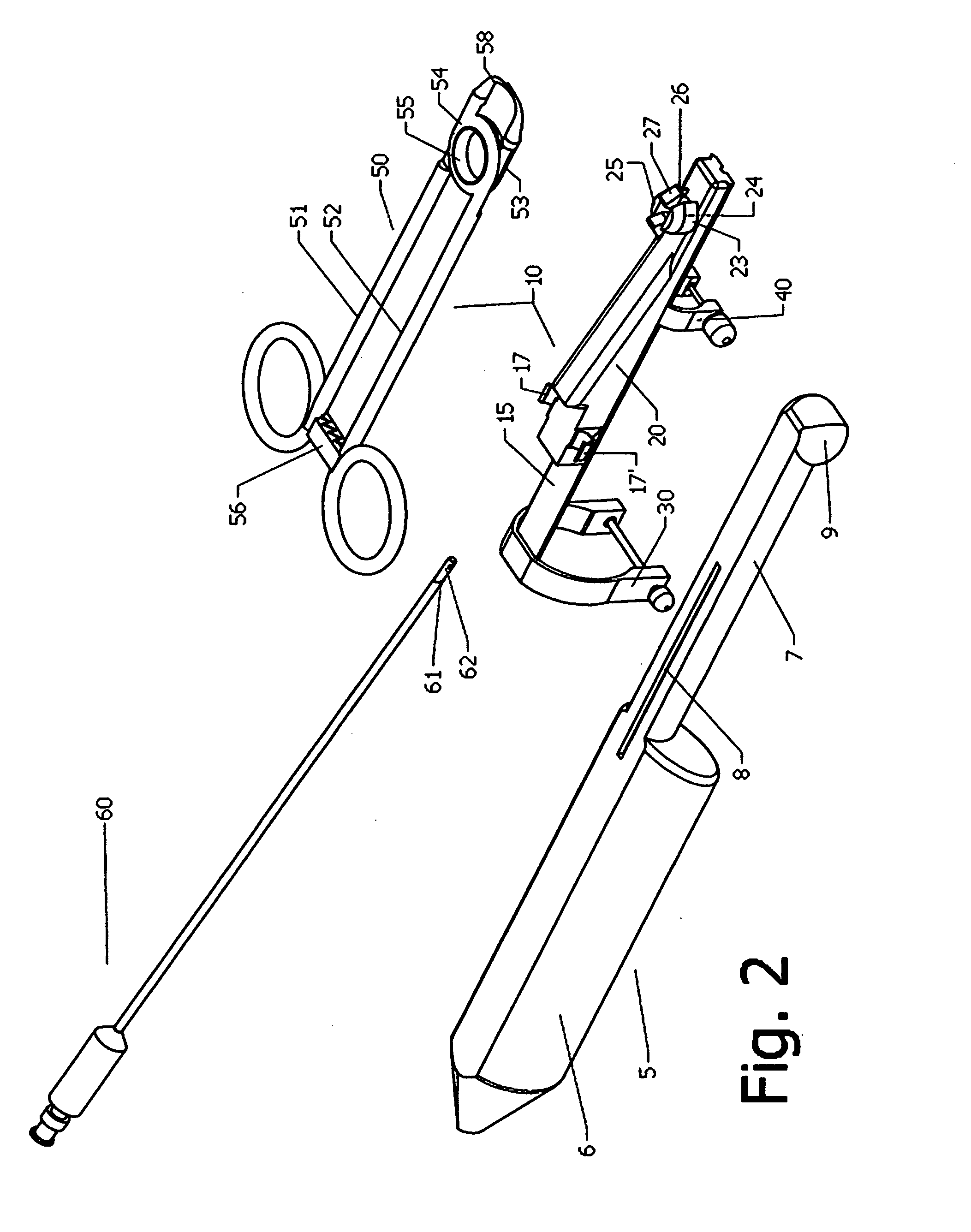
[0040]FIG. 1 presents an perspective right top view of the assembly 1, formed by the ultrasound probe 5 in combination with the puncture assistance device 10 to which the Veress needle 60 is added, and FIG. 2 presents an exploded view of the same. They depict an elongated ultrasound probe 5 that is otherwise known to be used for transvaginal or transrectal diagnostic procedures, with a flattened upper surface, formed of an elongated handle part 6 that serves to be gripped by the operator, an elongated shaft 7 that presents an elongated niche 8 on its flattened upper side (FIG. 2), and a rounded distal scanning part 9 that sends and receives ultrasound signals to / from the area to be examined that are converted to an image on t...


PUM


Abstract

A puncture assistance device for use in connection with an ultrasound probe to guide a Veress needle through the layers of the wall into a bodily cavity is provided. The puncture assistance device comprises a tenaculum-like forceps, a needle guide body provided with a slot for guiding the Veress needle within the scan plane of the ultrasound probe, attachment means to the ultrasound probe, and attachment means of the needle guide to the tenaculum-like forceps. Also provided is a balloon catheter for use as a retractor in gaining access into the abdominal cavity, presenting at its distal end an inflatable balloon membrane and an framework of non-distendable flexible fibers disposed in a specific arrangement. A method access the abdominal cavity with the aid of said devices under ultrasound guidance is also provided.

Description

PRIORITY CLAIM[0001]This invention claims priority to U.S. Provisional patent application Ser. No. 13 / 619,14997, filed on 12 Dec. 2013, and is incorporated by reference herein.BACKGROUND OF THE INVENTION[0002]Over the past 50 years, developments in electronic and optical technologies have meant that it has become possible to perform many operations laparoscopically. The unique feature distinguishing laparoscopic from open abdominal or vaginal surgery is the need to insert needles, trocars and cannulas for initial entry into the abdomen, the insertion of the primary trocar being made blindly in most cases. This may result in inadvertent bowel or vascular injury which can be responsible for major morbidity and mortality. In the last 25 years great efforts have been made for the prevention of these injuries by developing so called safe entry techniques especially because almost 50% of the complications of laparoscopic surgery occur at the time of primary entry site into the abdomen, be...

Claims


Application Information

Patent Timeline
IPC IPC(8): A61B17/34
CPCA61B17/3403A61B17/3439A61B17/3417A61B2017/3486A61B2017/3454A61M29/02A61M2029/025A61B2017/3413A61M25/10182A61B2017/3488A61B17/0281A61B17/3474A61B17/42A61B2017/00115A61B2017/00199A61B2017/00557A61B2017/320048A61B2017/4225A61B2090/378
Inventor ESANU, CATALIN
Owner ESANU CATALIN
Why Eureka
Try Eureka