Solid phase labeling method

a solid phase and labeling technology, applied in the field of protein labeling, can solve the problems of antibody loss during post-reaction purification steps, difficult to predict and additional, time-consuming steps, and achieve the effects of reducing antibody concentration, easy washing away, and controlling the degree of labeling

Inactive Publication Date: 2015-12-31
MU BIOTEKNIK
View PDF2 Cites 0 Cited by
  • Summary
  • Abstract
  • Description
  • Claims
  • Application Information

AI Technical Summary

Benefits of technology

[0009]A novel method for solid phase fluorescent labeling antibodies has been invented. By using a solid support throughout the labeling, excess reagents may be easily washed away while the antibody remains bound to the solid support. The solid support utilized in the inventive method comprises an affinity for an Fc portion of an antibody, e.g. a protein A affinity medium or a protein G affinity medium. Strong and specific interaction between such a solid support and antibodies, or fragments thereof retaining the Fc portion, in contrast to, e.g., the less specific interaction between an IMAC support and antibodies, allows for simultaneous purification, concentration and labeling of antibody samples. Since the solid support may be designed to interact with the Fc portion of various classes and subclasses of antibodies from a variety of species, the method need not be specifically adapted to different specific antibodies. Starting with 100-2000-fold lower antibody concentrations than what is required for labeling in solution, μg amounts of antibodies have been labeled with the inventive method. Further, the method provides a homogeneous labeling environment, which makes it possible to control the degree of labeling by adding different amounts of reactive dye, even if starting with impure antibody samples of low concentration, such as serum.

Problems solved by technology

The labeling reaction in solution is difficult to control and the resulting degree of labeling is hard to predict.
When the labeling reaction is finished, unreacted fluorescent label must be removed by dialysis or desalting columns adding one additional, time-consuming step.
Other disadvantages include dilution and loss of antibody during post-reaction purification steps as well as incomplete removal of free dye.
However, radiolabeling on protein A did not prove very effective, as the isotope incorporation was only 18% and 34% for 125I and ICI, respectively.
There is a great need for fluorescently labeled primary antibodies, but reproducibility problems, insufficient recovery of antibody and requirement of pure samples at a high concentrations often makes it impossible to obtain such antibodies using existing methods.

Method used


Image

  • Solid phase labeling method
  • Solid phase labeling method
  • Solid phase labeling method

Examples


Embodiment Construction

Materials and Methods

Materials

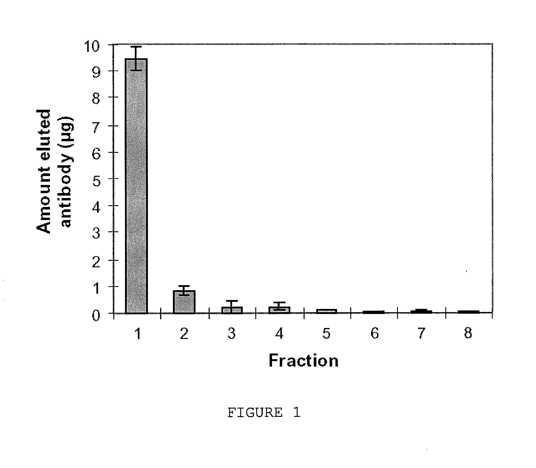
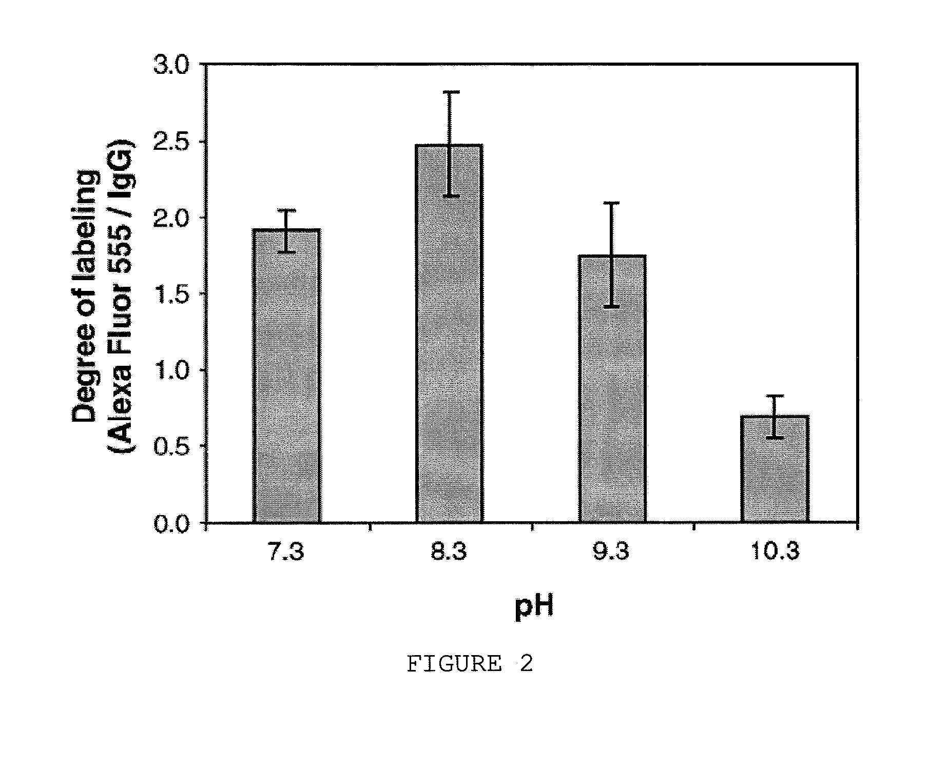
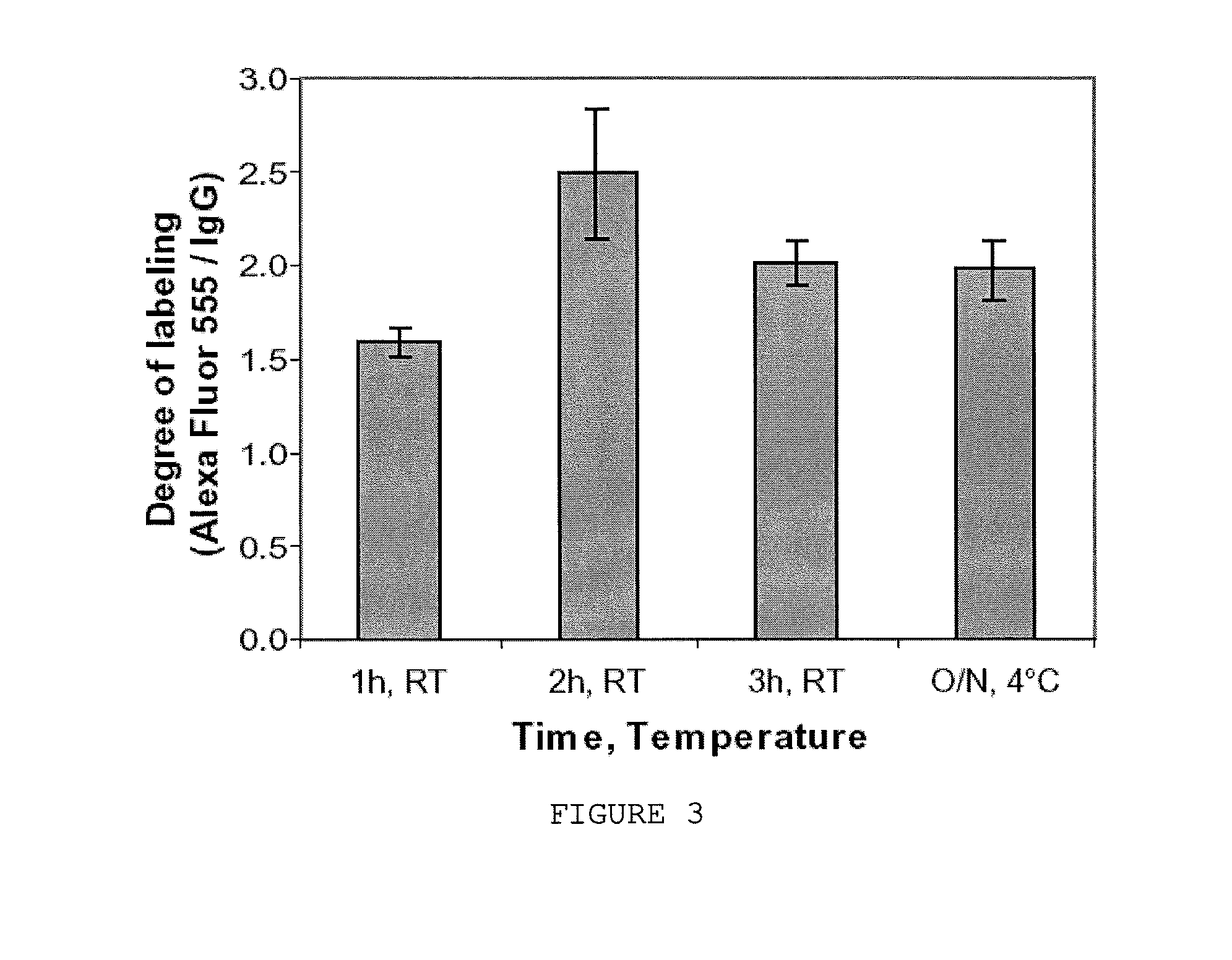
[0059]All chemicals were purchased from Sigma-Aldrich (Stockholm, Sweden) unless otherwise noted. The following Alexa Fluor® dyes were supplied by Invitrogen-Molecular Probes (Eugene, Oreg., US): succinimidyl ester of Alexa Fluor® 488 carboxylic acid (mixed isomers), succinimidyl ester of Alexa Fluor® 555 carboxylic acid and succinimidyl ester of Alexa Fluor® 647 carboxylic acid. 1 mg of each type of Alexa Fluor® dye was dissolved in DMSO to a final concentration of 40 μg / μl and stored at −80° C. Mono-specific polyclonal rabbit antibodies were obtained from the Human Protein Atlas program (http: / / www.proteinatlas.org / ). Antibodies used were α-His6-ABP (ABP refers to albumin binding protein), α-C1 tetrahydrofolate synthase, α-ornithine carbamoyltransferase, α-programmed cell death 4 isoform 1 and α-activator of 90 kDa heat shock protein ATPase homolog 1 (AHA1). Alexa Fluor® 555 rabbit anti-mouse IgG were obtained from Invitrogen-Molecular Probes (Eugene,...


PUM

PropertyMeasurementUnit
pHaaaaaaaaaa
concentrationaaaaaaaaaa

Abstract

The invention provides for the labeling of antibodies with a fluorescent label, using a solid support comprising an affinity for an Fc portion of an antibody. Thus, the invention provides a method for labeling an antibody or fragment thereof with a fluorescent label, comprising the steps of immobilizing an antibody on the solid support and covalently coupling a fluorescent label to the immobilized antibody, as well as a kit for performing such method.

Description

CROSS REFERENCE TO RELATED APPLICATIONS[0001]This application is a Continuation application of co-pending U.S. application Ser. No. 12 / 076,661 filed on Mar. 20, 2008, which claims priority under 35 U.S.C. 119(e) to U.S. Provisional Application No. 60 / 907,190 filed on Mar. 23, 2007, all of which are hereby expressly incorporated by reference into the present application.FIELD OF THE INVENTION[0002]The present invention relates to the field of labeling of proteins, in particular antibodies.BACKGROUND OF THE INVENTION[0003]Many applications in cell biology require use of antibodies for specific interactions and labeling of these antibodies with detector molecules for visualization. These detector molecules can, dependent on the application, be bioluminescent markers, enzymes or fluorescent labels. Fluorescently labeled antibodies are a very important tool in this field to provide for the specific detection of antigens. Fluorescently labeled antibodies are commonly used in immunohistoch...

Claims


Application Information

Patent Timeline
Patent Type & Authority Applications(United States)
IPC IPC(8): G01N33/533G01N33/548G01N33/545
CPCG01N33/533G01N33/545G01N33/548
Inventor UHLÉN, MATHIASANDERSSON SVAHN, HÉLÈNELUNDBERG, EMMA
Owner MU BIOTEKNIK
Why Eureka
Try Eureka