Systems and methods for detecting intracranial pressure and volume

a system and volume detection technology, applied in the field of systems and methods for detecting brain injuries, can solve the problems of significant impairment of a person's ability to function physically, cognitively and psychologically, increased risk of repeated head injuries in individuals with mtbi, and increased risk of brain damage to a second tim

Active Publication Date: 2016-01-07
AUGUSTA UNIV RES INST INC
View PDF5 Cites 19 Cited by
  • Summary
  • Abstract
  • Description
  • Claims
  • Application Information

AI Technical Summary

Benefits of technology

[0013]Still another embodiment provides a method and system for detecting distortions or deformations in the ONS that are indicative of trauma that occurred in the past and are also indicative of an increased risk of suffering brain injury from future trauma to the head.

Problems solved by technology

However tissue changes, as a result of MTBI at the neuronal level can lead to significant, life-long impairment of a person's ability to function physically, cognitively, and psychologically.
As a result of no diagnosis or the false belief that the initial injury has resolved, individuals with MTBI are at increased risk for repeated head injury, because they often do not avoid the activity that caused the initial concussion.
Because of the initial injury, physiological disturbances and disruption of the blood-brain barrier make the brain more susceptible to a second injury.
CTBI results in varying degrees of motor, cognitive and behavioral impairments.
However, neuropsychological testing requires specialized training to perform accurately, requires a substantial length of time to perform, and is not typically performed immediately after an injury.
These methods are still in development and are not practical to use at the point of injury.
Unfortunately, detection of optic nerve sheath diameter is difficult to perform automatically, and requires a skilled clinician to properly identify the appropriate nerve.
With limited diagnostic tools combined with the often subtle, mild symptoms, identification of this type of injury is difficult when suspected.

Method used


Image

  • Systems and methods for detecting intracranial pressure and volume
  • Systems and methods for detecting intracranial pressure and volume
  • Systems and methods for detecting intracranial pressure and volume

Examples


example 1

Cadaveric Trials

[0108]Materials and Methods

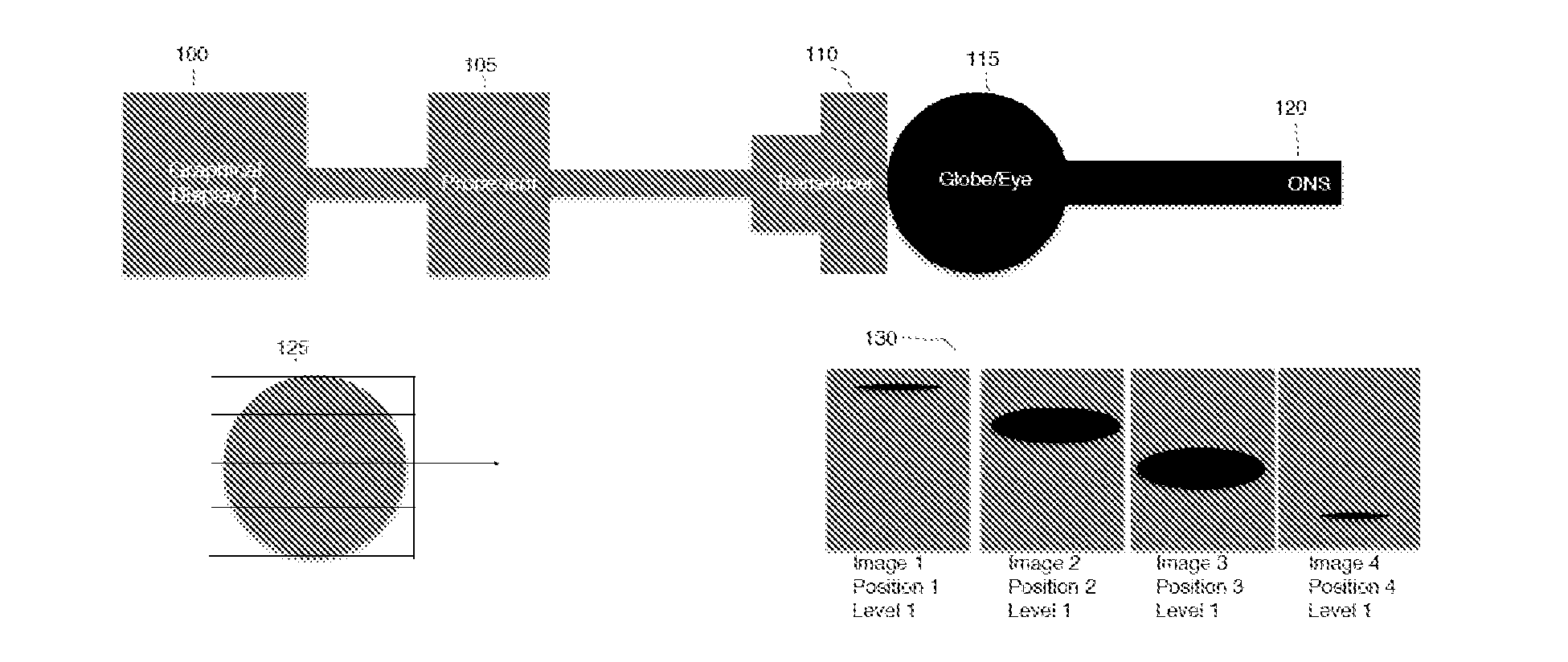
[0109]A cadaveric trial was chosen to study the speed at which the ONS dilates with increasing ICP. An invasive intracranial monitor was used to monitor the ICP as well as instill saline into the ventricle of the cadaver (increasing ICP) while simultaneous US scans of the ONS were performed to obtain 2D ONSD measurements.

[0110]Results

[0111]The ONS dilates near simultaneously with instillation of fluid into the intracranial space (ICS). The percent change in ONSD with change in ICP levels after saline instillation at different intervals. On an average, a 90% and 5% change in ICP and ONSD was recorded during the entire trial. Although the change in ONS dilation is simultaneous with ICP increase, the behavior of ONS dilation (varies from 4.2 mm to 5.3 mm) is less dynamic than change in ICP levels (4 cm / H2O to 34 cm / H2O). Therefore, there is an impetus to develop sensitive and multiple biomarkers than a traditional 2D ONSD to accurately detect ...

example 2

Human Trials

[0112]Materials and Methods

[0113]Data acquisition from 5 patients has been completed to date.

[0114]Trauma patients, who were both sedated and on mechanical ventilation, with recent brain injuries were potential subjects in this protocol. ICP measurements were obtained from an invasively placed intracranial monitor. 2D US measurements of the ONS were obtained using a SonoSite Micromaxx US system (L38 10-5 MHz linear transducer). 3D modeling of the ONS was made using a combination of the L38 transducer coupled to a sensor, which maps the position and orientation of the transducer during the US exam. The position and time-corrected US images were then automatically re-formatted to provide US data at any orthogonal or non-orthogonal slice plane. Using a 2½ D approach for surface reconstruction, a 3D surface model of the ONS was generated from the segmented boundary slices. The ONSD measurements derived from a 2D image and from the 3D model of the ONS were compared to the dir...

example 3

3D Modeling of the Optic Nerve Sheath (ONS)

[0117]Materials and Methods

[0118]A 2D ultrasound transducer was used according to the manufacturer's instructions. The MetriTrack™ ultrasound system was used. This approach allows minimal-overhead 3D scanning with an innovative add-on device that is clipped onto standard ultra sound probes, near-instantaneous calibration, and has the potential to guide a variety of other image-guided interventions. Current version of CGM software version will perform “hands-off” 3D ultrasound reconstruction without system / operator interaction (FIGS. 6A and 6B). This probe tracking and reconstruction is performed with sensors entirely integrated into a clip-on bracket for handheld US probes (FIGS. 6A and 6B), with stereo cameras observing the environment (both on the patient and the surrounding background) and extract features that are salient enough to support probe trajectory reconstruction. Furthermore, in adverse conditions 3D environment surfaces can be...


PUM


Abstract

It has been discovered that even mild changes in cerebrospinal fluid (CSF) pressure or intracranial pressure (ICP) can be detected immediately as evidenced by distortions in the ONS surface structure. Further, the changes in the ONS persist after the CSF pressure has returned to normal. The stability of ONS distortions provides a means of detecting transient changes in brain pressure even when the use of the diagnostic ultrasound is delayed. One embodiment provides systems and methods for detecting or diagnosing brain injury by detecting distortions or deformations of the ONS, preferably using ultrasound.

Description

CROSS REFERENCE TO RELATED APPLICATIONS[0001]This application claims benefit of and priority to U.S. Provisional Patent Application No. 62 / 019,624 filed on Jul. 1, 2014, and is incorporated by reference in its entirety.FIELD OF THE INVENTION[0002]The invention is generally directed to systems and methods for detecting brain injury in a subject.BACKGROUND OF THE INVENTION[0003]According to the Center of Disease Control and Prevention, an estimated 1.7 million people sustain a traumatic brain injury (TBI) annually (Faul M, Xu L, Wald M M, Coronado V G. Traumatic Brain Injury in the United States: Emergency Department Visits, Hospitalizations and Deaths 2002-2006. Atlanta (GA): Centers for Disease Control and Prevention, National Center for Injury Prevention and Control; 2010.). Out of that 1.7 million people, 52,000 die, 275,000 are hospitalized, and 1.365 million are treated and released from an emergency department.[0004]TBI is a spectrum, from mild, almost unnoticeable brain injury...

Claims


Application Information

Patent Timeline
Patent Type & Authority Applications(United States)
IPC IPC(8): A61B5/00A61B5/03A61B5/055A61B6/03A61B8/00A61B8/08A61B8/10
CPCA61B5/055A61B6/032A61B6/037A61B8/10A61B8/4254A61B5/031A61B8/461A61B8/5207A61B5/4058A61B5/004A61B8/483A61B5/684A61B5/7282A61B5/6821A61B2576/02A61B5/0035A61B8/4494A61B6/506A61B6/5217A61B8/14A61B8/463A61B8/488A61B8/5223A61B8/5246A61B6/501G16H50/30
Inventor LYON, MATTHEW
Owner AUGUSTA UNIV RES INST INC
Why Eureka
Try Eureka