Use of lifr or fgfr3 as a cell surface marker for isolating human cardiac ventricular progenitor cells

a cell surface marker and human heart technology, applied in 3d culture, skeletal/connective tissue cells, instruments, etc., can solve the problems of unsuitable use of a large quantity of viable cells, unmet needs, etc., to enhance ventricular function, improve the function of a damaged heart, and easy and rapid cell isolation

Active Publication Date: 2016-04-21
PROCELLA THERAPEUTICS AB
View PDF3 Cites 16 Cited by
  • Summary
  • Abstract
  • Description
  • Claims
  • Application Information

AI Technical Summary

Benefits of technology

[0011]Transplantation of the ventricular progenitor cells provided herein produces a pure, functional and mature human ventricular muscle organ of large size (e.g., twice the size of the murine heart) that can generate force, respond to catecholamines, lose automaticity, contain T tubules and display hypertrophic growth of adult rod-shaped cells by 5 months post-transplantation. Thus, human ventriculogenesis can be achieved via a cell autonomous pathway driven by the purified ISL1/LIFR/FGFR3 human ventricular progenitors provided herein. These HPVs provided herein allow for new in vivo models of human cardiac disease in murine-human chimeras and for the development of organ-on-organ regenerative therapeutic strategies for cardiac disease.
[0012]This identification of a key cell surface marker of cardiac ventricular progenitor cells allows for easy and rapid isolation of the cells. Furthermore, determination of culture conditions for expansion and ventricular lineage bias of the cells allows for the preparation of large cultures (a billion or more cells) of a clonal population of cardiac ventricular progenitor cells. These cells can be used, for example, to improve function in a damaged heart in a subject, particularly damage in the ventricular region. The progenitor cells can be transplanted in vivo for dif

Problems solved by technology

Although heart transplantation can be curative, the markedly limited availability of human heart organ donors has led to a widespread unmet clinical need for a renewable source of pure, mature and functional human ventricular muscle tissue (Segers, V. F. M. and Lee, R. J. (2008) Nature 451:937-942; Spater, D. et al.
While human pluripotent stem cells hold great promise, a significant challenge has been the ability to move from simply d

Method used


Image

  • Use of lifr or fgfr3 as a cell surface marker for isolating human cardiac ventricular progenitor cells
  • Use of lifr or fgfr3 as a cell surface marker for isolating human cardiac ventricular progenitor cells
  • Use of lifr or fgfr3 as a cell surface marker for isolating human cardiac ventricular progenitor cells

Examples


example 1

Generation of Human Isl1+ Cardiomyogenic Progenitor Cells by Modulation of Wnt Signaling in Human Pluripotent Stem Cells

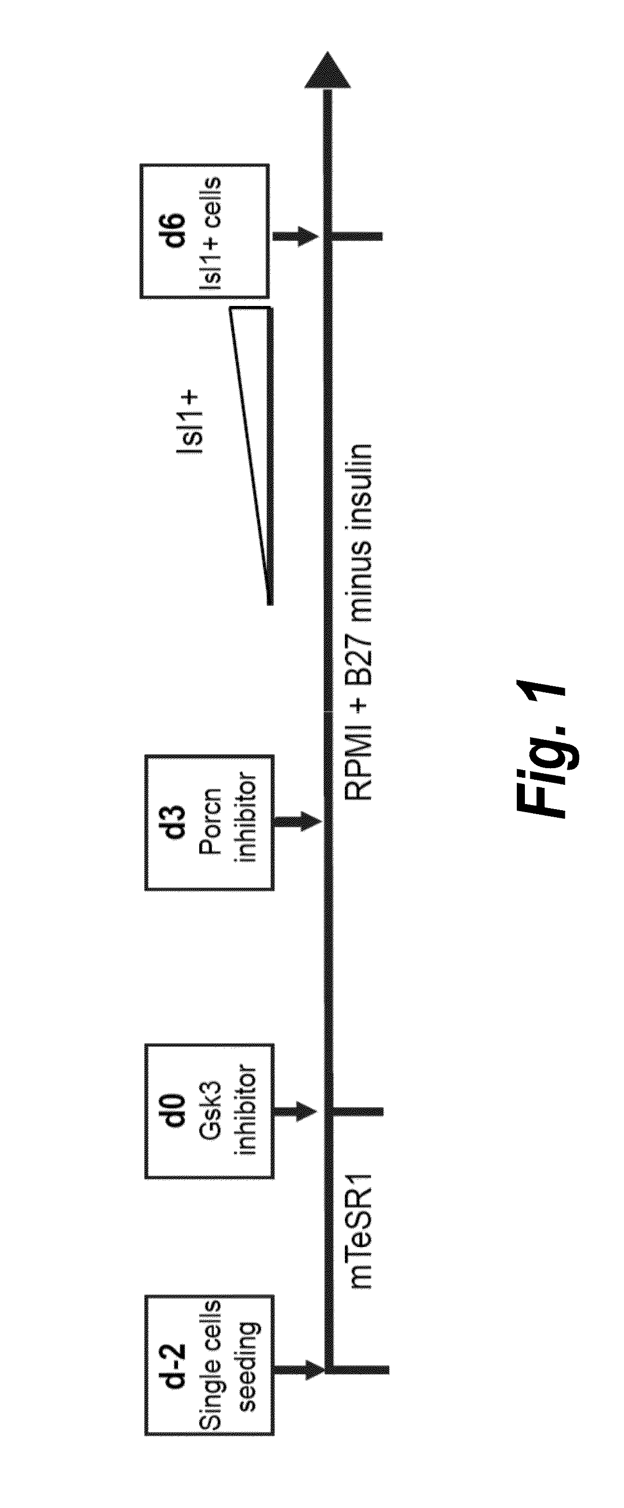
[0206]Temporal modulation of canonical Wnt signaling has been shown to be sufficient to generate functional cardiomyocytes at high yield and purity from numerous hPSC lines (Lian, X. et al. (2012) Proc. Natl. Acad. Sci. USA 109:E1848-1857; Lian, X. et al. (2013) Nat. Protoc. 8:162-175). In this approach, Wnt / β-catenin signaling first is activated in the hPSCs, followed by an incubation period, followed by inhibition of Wnt / β-catenin signaling. In the originally published protocol, Wnt / β-catenin signaling activation was achieved by incubation with the Gsk3 inhibitor CHIR99021 (GSK-3 α, IC50=10 nM; GSK-3, β IC50=6.7 nM) and Wnt / β-catenin signaling inhibition was achieved by incubation with the Porcn inhibitor IWP2 (IC50=27 nM). Because we used Gsk3 inhibitor and Wnt production inhibitor for cardiac differentiation, this protocol was termed GiWi protocol. To improve t...

example 2

Identification of Jagged 1 as a Cell Surface Marker of Cardiac Progenitor Cells

[0214]To profile the transcriptional changes that occur during the cardiac differentiation process at a genome-scale level, RNA sequencing (RNA-seq) was performed at different time points following differentiation to build cardiac development transcriptional landscapes. We performed RNA-seq experiments on day 0 to day 7 samples, as well as day 19 and day 35 samples (two independent biological replicates per time point). Two batches of RNA-seq (100 bp and 50 bp read length) were performed using the illumine Hiseq 2000 platform. In total, 20 samples were examined. Bowtie and Tophat were used to map our reads into a reference human genome (hg19) and we calculate each gene expression (annotation of the genes according to Refseq) using RPKM method (Reads per kilobase transcript per million reads). Differentiation of hPSCs to cardiomyocytes involves five major cell types: pluripotent stem cells (day 0), mesoder...

example 3

Clonal Differentiation of Isl1+Jag1+ Cardiac Progenitor Cells

[0223]To characterize the clonal differentiation potential of Isl1+Jag1+ cells, cardiomyogenic progenitor cells were generated by the culturing protocol described in Example 1, and one single Isl1+Jag1+ cell was seeded into one well of a Matrigel-coated 48-well plate. Cells were purified with antibody of Jag1 and then one single cell was seeded into one well. The single cells were then cultured for 3 weeks in Cardiac Progenitor Culture (CPC) medium (advanced DMEM / F12 supplemented with 2.5 mM GlutaMAX, 100 μg / ml Vitamin C, 20% Knockout Serum Replacement).

[0224]Immunostaining of the 3-week differentiation cell population was then performed with three antibodies: cardiac troponin I (cTn1) for cardiomyocytes, CD144 (VE-cadherin) for endothelial cells and smooth muscle actin (SMA) for smooth muscle cells. The results showed that the single cell-cultured, Isl1+Jag1+ cells gave rise to cTnI positive and SMA positive cells, but no...


PUM


Abstract

The present invention provides LIFR and FGFR3 as cell surface markers for isolating human cardiomyogenic ventricular progenitor cells, in particular progenitor cells that preferentially differentiate into cardiac ventricular muscle cells. Thus, the invention provides human ventricular progenitor (HVP) cells. The invention provides in vitro methods of the separation of Islet 1+ LIFR+ ventricular progenitor cells and/or Islet 1+/FGFR3+ ventricular progenitor cells and/or Islet 1+/LIFR+/FGFR3+ ventricular progenitor cells, and the large scale expansion and propagation thereof. Large clonal populations of isolated LIFR+ and/or FGFR3+ ventricular progenitor cells are also provided. Methods of in vivo use of LIFR+ and/or FGFR3+ ventricular progenitor cells for cardiac repair or to improve cardiac function are also provided. Methods of using the LIFR+ and/or FGFR3+ ventricular progenitor cells for cardiac toxicity screening of test compounds are also provided.

Description

RELATED APPLICATIONS[0001]This application is a continuation in part of U.S. Ser. No. 14 / 832,324, filed on Aug. 21, 2015, which claims the benefit of the priority date of U.S. Provisional Application No. 62 / 040,892, filed on Aug. 22, 2014, and U.S. Provisional Application No. 62 / 194,016, filed on Jul. 17, 2015. The content of these applications is hereby incorporated by reference in its entirety.BACKGROUND OF THE INVENTION[0002]Heart failure, predominantly caused by myocardial infarction, is the leading cause of death in both adults and children worldwide and is increasing exponentially worldwide (Bui, A. L. et al. (2011) Nat. Rev. Cardiol. 8:30-41). The disease is primarily driven by the loss of ventricular muscle that occurs during myocardial injury (Lin, Z. and Pu, W. T. (2014) Sci. Transl. Med. 6:239rv1) and is compounded by the negligible ability of the adult heart to mount a regenerative response (Bergmann, O. et al. (2009) Science 324:98-102; Senyo, S. E. et al. (2013) Nature...

Claims


Application Information

Patent Timeline
IPC IPC(8): C12N5/0775A61K35/34G01N33/50
CPCC12N5/0662G01N33/5014C12N2501/235C12N2501/10A61K35/34C12N5/0657C12N2501/119C12N2501/415C12N2501/42C12N2506/02C12N2506/45C12N2513/00
Inventor CHIEN, KENNETH R.LIAN, XIAOJUN LANCE
Owner PROCELLA THERAPEUTICS AB
Why Eureka
Try Eureka