

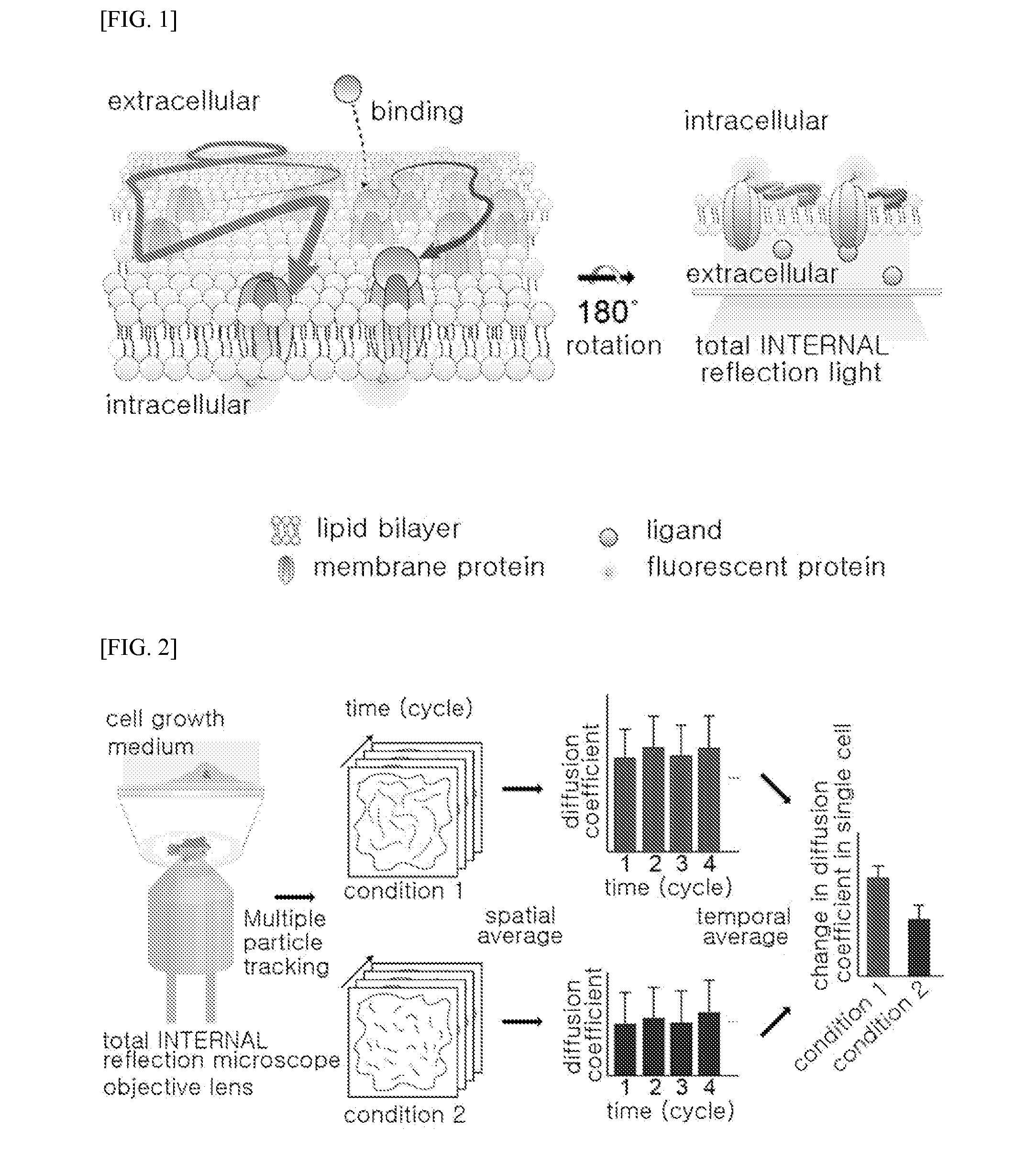
Method of analyzing binding aspect of membrane protein in a living cell
a technology of membrane protein and binding aspect, which is applied in the field of living cell binding aspect analysis, can solve the problems of limited analysis of the interaction between and it is fundamentally difficult to measure the binding of ligands to the extracellular domain of the target membrane protein, etc., and achieves rapid and easy analysis of the binding aspect, accurate, sensitive
- Summary
- Abstract
- Description
- Claims
- Application Information
AI Technical Summary
Benefits of technology
Problems solved by technology
Method used
Image

Examples
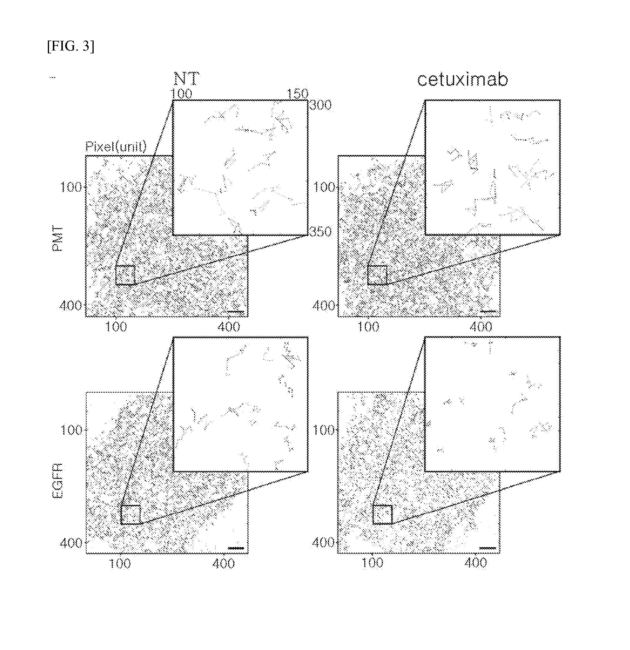
example 1
Measurement of Target Membrane Protein: EGFR
[0093]1.1 Plasmid for Preparing EGFR-mEos3.2 Fusion Protein
[0094]EGFR WT (Addgene plasmid #11011) used in the experiment was provided by Dr. Matthew Meyerson, and mEos3.2 fluorescent protein was provided by Dr. Tao xu. The given fluorescent protein was subcloned from pEGFP-N1 / mEos3.2 to form pcDNA3.1 / mEos3.2-his in order to facilitate binding with a membrane protein. To prepare a protein in which the membrane protein EGFR WT (SEQ ID NO: 1) is linked with the fluorescent protein mEos3.2 (SEQ ID NO: 2), pcDNA3.1 / EGFR WT-mEos3.2-His was constructed using restriction enzymes such as Xbal (SEQ ID NO: 3, TCTAGA) and NotI (SEQ ID NO: 4, GCGGCCGC).
[0095]1.2 Plasmid for Preparing PMT-mEos3.2 Fusion Protein
[0096]A plasmid in which PMT is linked with a fluorescent protein was prepared to be used as a control group for Example 1.1. DNA (SEQ ID NO: 5) corresponding to PMT was inserted into the subcloned pcDNA3.1 / mEos3.2-his using restriction enzymes su...
example 2
Measurement of Target Membrane Protein: GPCR
[0112]2.1 Plasmid for Preparing β2-AR-mEos3.2 Fusion Protein
[0113]β2-AR (Addgene plasmid #14697) used in the experiment was provided by Dr. Robert Lefkowitz, and β2-AR DNA (SEQ ID NO: 6) was inserted into the pcDNA3.1 / mEos3.2-his subcloned in Example 1.1 using restriction enzymes such as BamHI and Xbal, thereby constructing pcDNA3.1 / β2-AR-mEos3.2-His.
[0114]2.2 Plasmid for Preparing FZD1-mEos3 Fusion Protein
[0115]FZD1 (Addgene plasmid #16819) used in the experiment was provided by Dr. Randall Moon, and FZD1 DNA (SEQ ID NO: 7) was inserted into the pcDNA3.1 / mEos3.2-his subcloned in Example 1.1 using restriction enzymes such as BamHI and Xbal, thereby constructing pcDNA3.1 / FZD1-mEos3.2-His.
[0116]2.3 Preparation of Host Cells for Expressing Fusion Protein
[0117]For an experiment to inactivate GPCR, COS7 cells were transfected with J32-AR-mEos3.2, FZD1-mEos3.2, and PMT-mEos3.2 (refer to Example 1.2) by the same method as described in Example 1.3...
example 3
Measurement of Target Membrane Protein in Different Environments
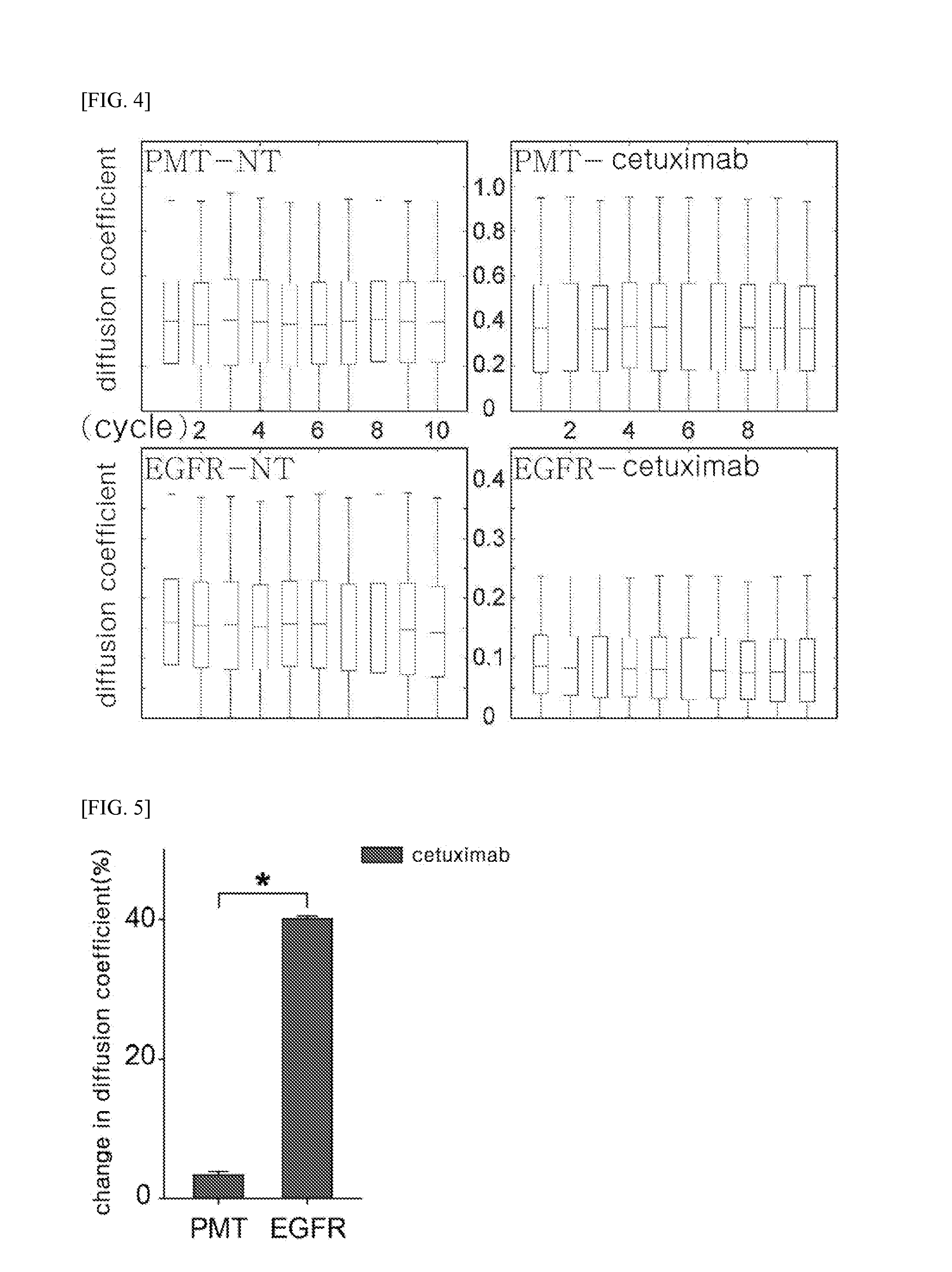
[0124]To prove technical reproducibility of the method described in Example 1, three groups of transfected COS7 cells were prepared on different days (batch#1, batch#2, and batch#3), two groups of transfected COS7 cells (batch#3-1 and batch#3-2) were prepared independently but on the same day, and then coverslips on which these batches were seeded were prepared. The host cells were treated with cetuximab by the same method as described in Example 1.4, and then a change in diffusion coefficient of EGFR was measured, and the results are shown in FIG. 9.
[0125]FIG. 9 illustrates the changes in diffusion coefficient of EGFR measured by the above-described method, in which batch#1, batch#2, and batch#3 represent the changes in diffusion coefficient of EGFR in the COS7 cells prepared in three different batches, and batch#3-1 and batch#3-2 represent changes in diffusion coefficient of EGFR measured at different glasses, but in ...
PUM
| Property | Measurement | Unit |
|---|---|---|
| Fraction | aaaaa | aaaaa |
| Molecular weight | aaaaa | aaaaa |
| Fluorescence | aaaaa | aaaaa |
Abstract
Description
Claims
Application Information