Eureka AIR delivers breakthrough ideas for toughest innovation challenges, trusted by R&D personnel around the world.

Ultrasound-Mediated Inducement, Detection, and Enhancement of Stable Cavitation

Active Publication Date: 2016-05-26
UNIVERSITY OF CINCINNATI
View PDF3 Cites 0 Cited by
  • Summary
  • Abstract
  • Description
  • Claims
  • Application Information

AI Technical Summary

Benefits of technology

This patent describes a method to improve sonothrombolysis treatment by using a nucleating agent and a thrombolytic agent in combination with ultrasonic energy. The ultrasonic energy is provided in intervals, with no energy during rest periods. This method enhances stable cavitation during sonothrombolysis, resulting in improved treatment outcomes.

Problems solved by technology

Ischemic strokes can lead to a variety of physical complications including permanent neurological damage and death.
When brain tissue is deprived of oxygen for more than 60-90 seconds, the brain tissue loses its function; when brain tissue is deprived of oxygen for greater than three hours, irreversible injury results, leading to infarction.
Currently, treatment of ischemic stroke is generally limited to thrombolytic therapies, whereby a blood clot is broken up or dissolved.
However, this therapy possesses a number of drawbacks.
Further, there is a 6.4% incidence of intracerebral hemorrhage in patients receiving this thrombolytic therapy.
However, these detection methods are also limited.
Further, detection methods have yet to be employed to enhance stable cavitation during sonothrombolysis.

Method used


Image

  • Ultrasound-Mediated Inducement, Detection, and Enhancement of Stable Cavitation
  • Ultrasound-Mediated Inducement, Detection, and Enhancement of Stable Cavitation
  • Ultrasound-Mediated Inducement, Detection, and Enhancement of Stable Cavitation

Examples


example 1

Passive Cavitation Detection with Dual Element Annular Array

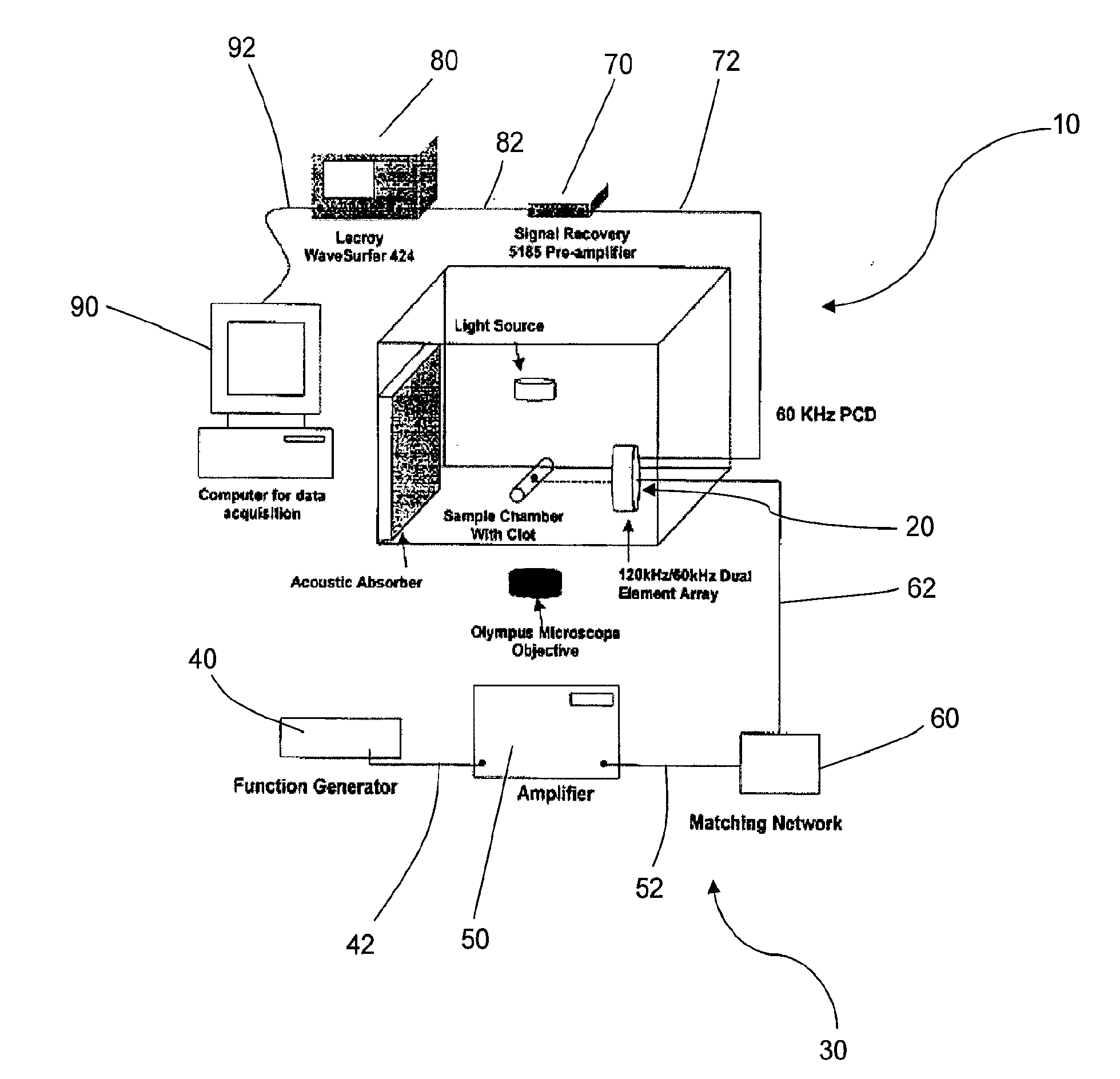
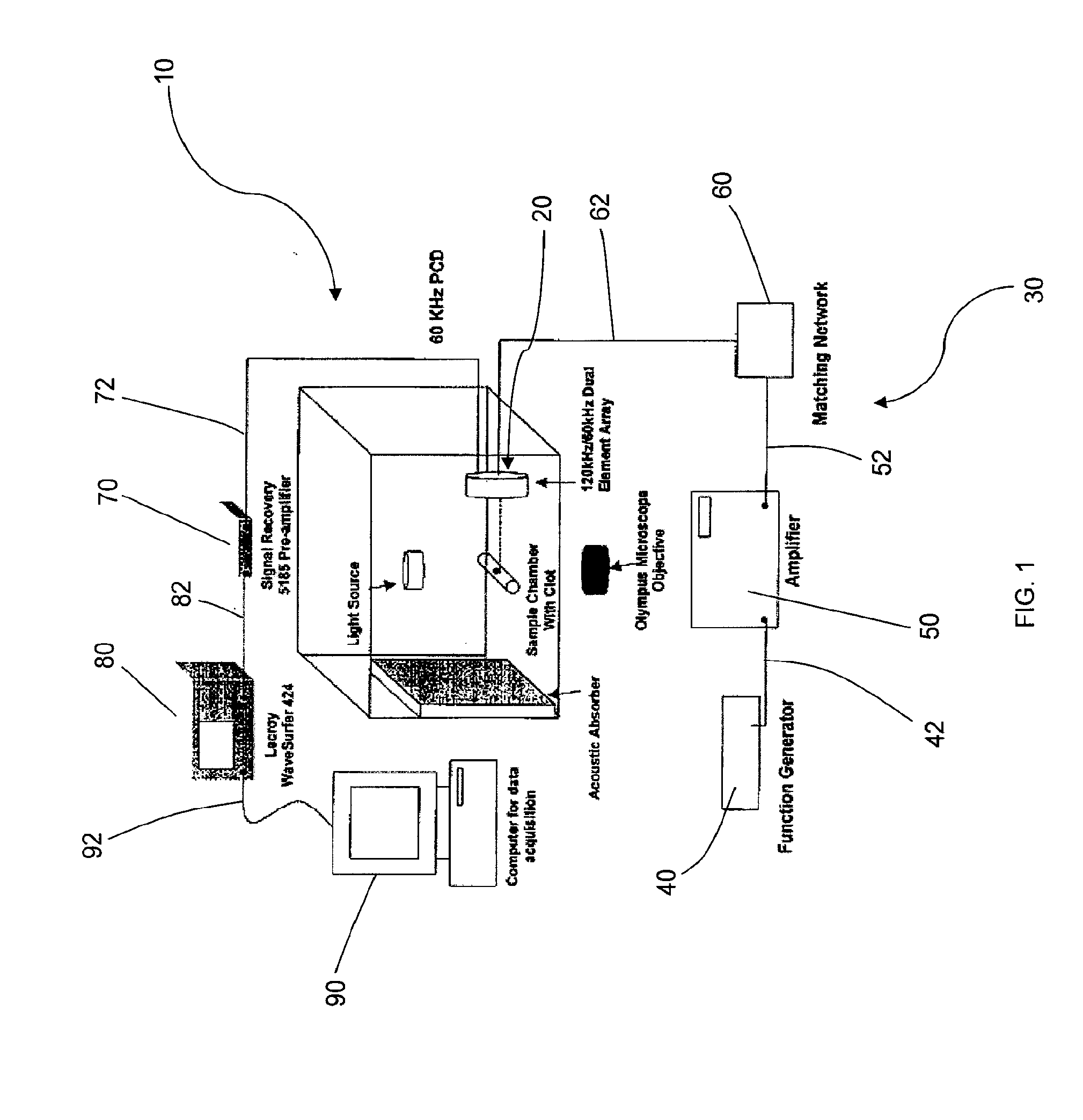
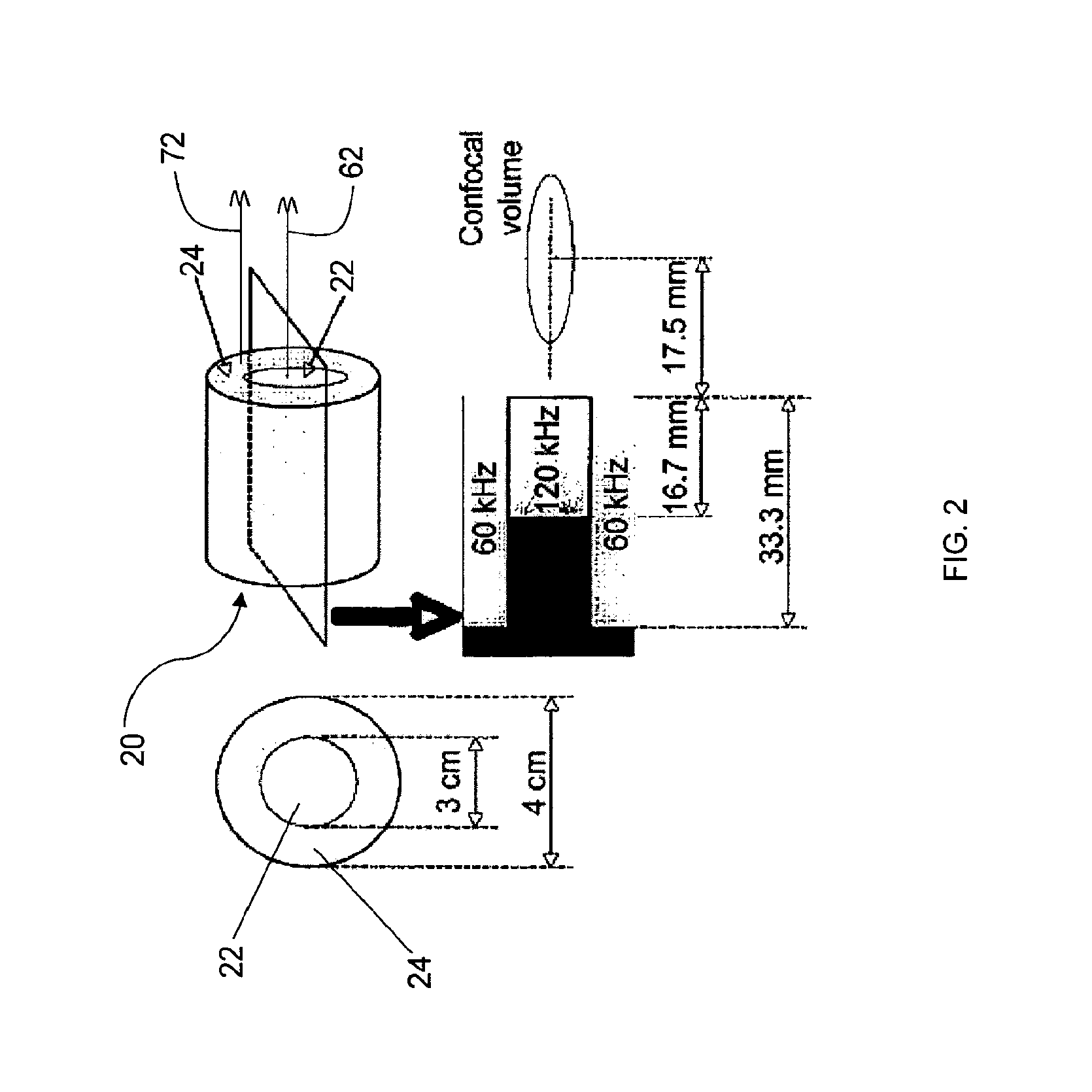
[0098]Dual Element Annular Array for 120-kHz Sonothrombolysis and 60-kHz Passive Cavitation Detection. A dual element annular array (FIG. 1) was designed to enable inducing and passively detecting stable cavitation during sonothrombolysis. To test the feasibility of this design approach, acoustic radiation from the 3 cm, 120-kHz source was computed using an exact series solution for the field of a baffled circular radiator in a homogeneous medium. Using the same method, the spatial sensitivity pattern of the surrounding annular passive cavitation detector (inner diameter 3 cm, outer diameter 4 cm) was computed at the subharmonic frequency of 60 kHz. Cross sections of the beam patterns are shown in FIG. 6. The field of the 120-kHz source had a −6 dB depth of field of 46 mm and a −6 dB beamwidth of 1.4 cm. The annular broadband passive cavitation detector had a collimated beam with amplitude 0.84 (−1.5 dB relative to surface ...

example 2

Effects of Stable Cavitation on Thrombolysis

[0103]Cavitation Nucleation with Infusion of Contrast Agent in an In Vitro Human Clot Model. An approach for inducing cavitation using infusion of a contrast agent, Definity®, was tested experimentally in vitro. Human whole blood clots and rt-PA (96 m / ml) were placed in human fresh frozen plasma in a thin-walled latex sample holder which was placed in a tank of water at 37° C. Percent clot mass loss was assessed as a function of peak-to-peak acoustic pressure for the following treatments: (a) no rt-PA, no Definity®, no pulsed ultrasound (the control); (b) rt-PA alone; (c) rt-PA, Definity® infusions, and pulsed ultrasound (˜0.12 MPa peak-to-peak pressure amplitude); (d) rt-PA, Definity® infusions, and pulsed ultrasound (˜0.21 MPa peak-to-peak pressure amplitude); (e) no rt-PA, Definity® infusions, and pulsed ultrasound (˜0.32 MPa peak-to-peak pressure amplitude); (f) rt-PA, phosphate buffered saline infusions (no Definity®) and pulsed ultra...

example 3

Effects of Stable Cavitation on Thrombolysis in an Ex Vivo Porcine Artery Model with Interval Ultrasound

[0110]Ex Vivo Porcine Carotid Artery Model with Physiologic Flows and Pressures. Porcine whole blood clots were inserted into living, excised porcine carotid arteries, and kept viable in a thin-walled latex chamber filled with degassed artificial cerebrospinal fluid while oxygenated plasma flowed through the lumen. The chamber was placed in a tank containing degassed filtered water at 37° C. A series of experiments were performed by infusing 1 ml / min plasma with 0.31 μl Definity® per 1 ml plasma through a porcine artery filled with a porcine whole blood clot at a physiologic pressure of 100±15 mmHg. Each clot and artery were insonated with 120 kHz continuous wave ultrasound at peak-to-peak pressures ranging from 0.37 MPa to 0.54 MPa for 45 seconds. The signals were analyzed for stable and inertial cavitation power.

[0111]Ultrasound Source Transducer and Passive Cavitation Detector....


PUM


Abstract

Methods and systems for passively detecting stable cavitation and enhancing stable cavitation during sonothrombolysis are provided. The method of passively detecting stable cavitation includes providing a determined level of ultrasonic energy and detecting a scattered level of ultrasonic energy. The system for inducing and passively detecting stable cavitation includes a dual-element annular transducer array configured to provide a fundamental ultrasonic frequency and to detect an ultrasonic frequency that is a derivative of the fundamental frequency. The method of enhancing stable cavitation includes administering a nucleating agent and a thrombolytic agent to a treatment zone, providing a determined level of ultrasonic energy, and detecting a scattered level of ultrasonic energy.

Description

[0001]This application is a divisional application of U.S. application Ser. No. 13 / 257,657, filed on Feb. 27, 2012, which claims priority to Int'l Application No. PCT / US2010 / 027992, filed on Mar. 19, 2010, which claims priority to U.S. Provisional Application Ser. No. 61 / 162,061, filed Mar. 20, 2009, the contents of which are hereby incorporated by reference in their entirety.TECHNICAL FIELD[0002]The present invention relates to methods and systems of inducing, detecting, and enhancing stable cavitation using ultrasound. More specifically, the present invention relates to methods and systems of inducing, passively detecting, and enhancing stable cavitation during sonothrombolysis.BACKGROUND[0003]Due to the prevalence of thrombo-occlusive disease worldwide and the need for improved clinical treatments, ultrasound has been investigated, either alone or in combination with thrombolytic drugs, to improve recanalization in patients with this disease. A common thrombo-occlusive disease is...

Claims


Application Information

Patent Timeline
IPC IPC(8): A61N7/00
CPCA61N2007/0039A61N7/00A61B8/06A61B8/481G01S7/52038A61B8/0808A61M37/0092A61B2017/22008A61B2017/22088A61B17/2202A61B2017/00172A61B2017/0019A61B2017/00194
Inventor HOLLAND, CHRISTY K.DATTA, SAURABHMAST, T. DOUGLASIVANCEVICH, NIKOLASHITCHCOCK, KATHRYN ELIZABETHHAWORTH, KEVIN
Owner UNIVERSITY OF CINCINNATI
Why Eureka
Eureka Blog
Learn More