Head fixing device for radiography imaging and x-ray imaging system having same

a technology of x-ray imaging and fixing device, which is applied in the direction of radiation diagnostics for dentistry, medical science, diagnostics, etc., can solve the problems of radiographs being difficult to use as accurate image data, deformation of facial contours in radiographs, and change of facial soft tissues, so as to minimize or prevent deformation of facial contours, minimize or prevent the effect of error

Inactive Publication Date: 2016-06-30
UNIV IND COOP GRP OF KYUNG HEE UNIV
View PDF5 Cites 7 Cited by
  • Summary
  • Abstract
  • Description
  • Claims
  • Application Information

AI Technical Summary

Benefits of technology

[0035]A head fixing apparatus for radiography, i.e. a head fixing apparatus for holding the head of a subject in a radiography position during radiography provides the following effects.
[0036]According to the present invention, the head fixing apparatus allows the head of a subject to be stably held in position during general radiography and dental radiography for the head, thereby minimizing a risk of error during radiography and providing an image having high diagnostic value.
[0037]In addition, according to the present invention, the head fixing apparatus can minimize or prevent deformation of a facial contour caused by compressi

Problems solved by technology

However, such a typical image processing system has the following problems in acquisition of an x-ray image and a facial appearance image and registration between the images.
If radiography is performed to obtain images of a skull, teeth, or a jawbone while a subject supports his/her chin on a chin rest, soft tissues of the chin are pressed by the chin rest, causing change of facial soft tissues, which causes deformation of a facial contour in a radiograph.
Such a radiograph is difficult to use as accurate image data when it is important to obtain the contour of facial soft tissues as well as conditions of hard tissues.
In other words, since the chin is pressed by the chin rest to cause change of facial soft tissues, the degree of registration between the facial appearance image and the x-ray image is reduced, thereby making acquisition of accurate diagnostic data difficult.
In addition, in order to increase the degree of registration between the facial appearance image and the x-ray image, additional processes such as compensation of the x-ray image and/or the facial appearance image are required, thereby causing unnecessary consumption of time and manpower.
However, a radiograph in which facial soft tissues (face skin) is deformed by compression of a skin of a mental region cannot serve as accurate diagnostic data in a procedure requiring consideration of appearance.
In addition, since a contour of soft tissues in a radiograph

Method used


Image

  • Head fixing device for radiography imaging and x-ray imaging system having same
  • Head fixing device for radiography imaging and x-ray imaging system having same
  • Head fixing device for radiography imaging and x-ray imaging system having same

Examples


Embodiment Construction

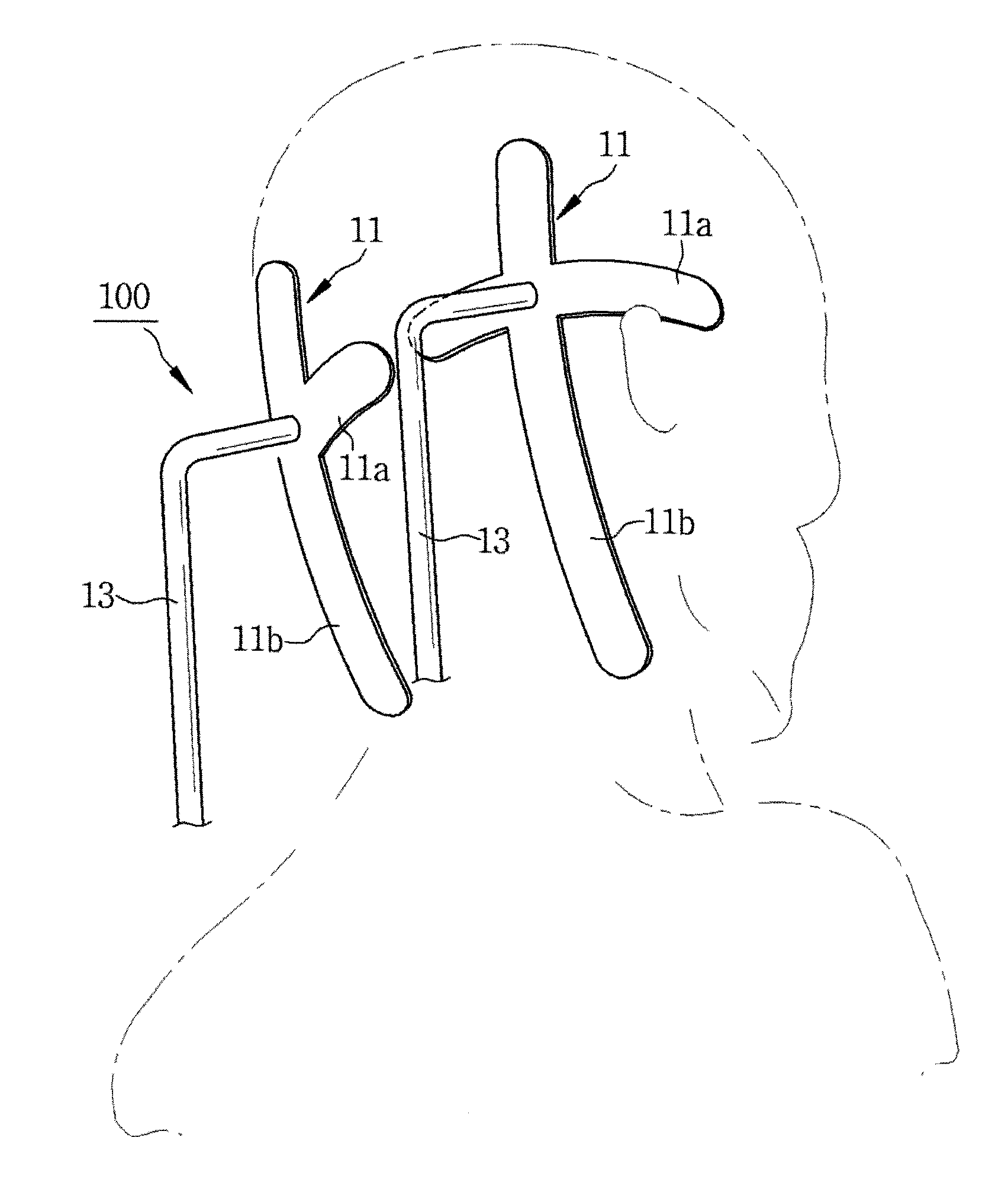
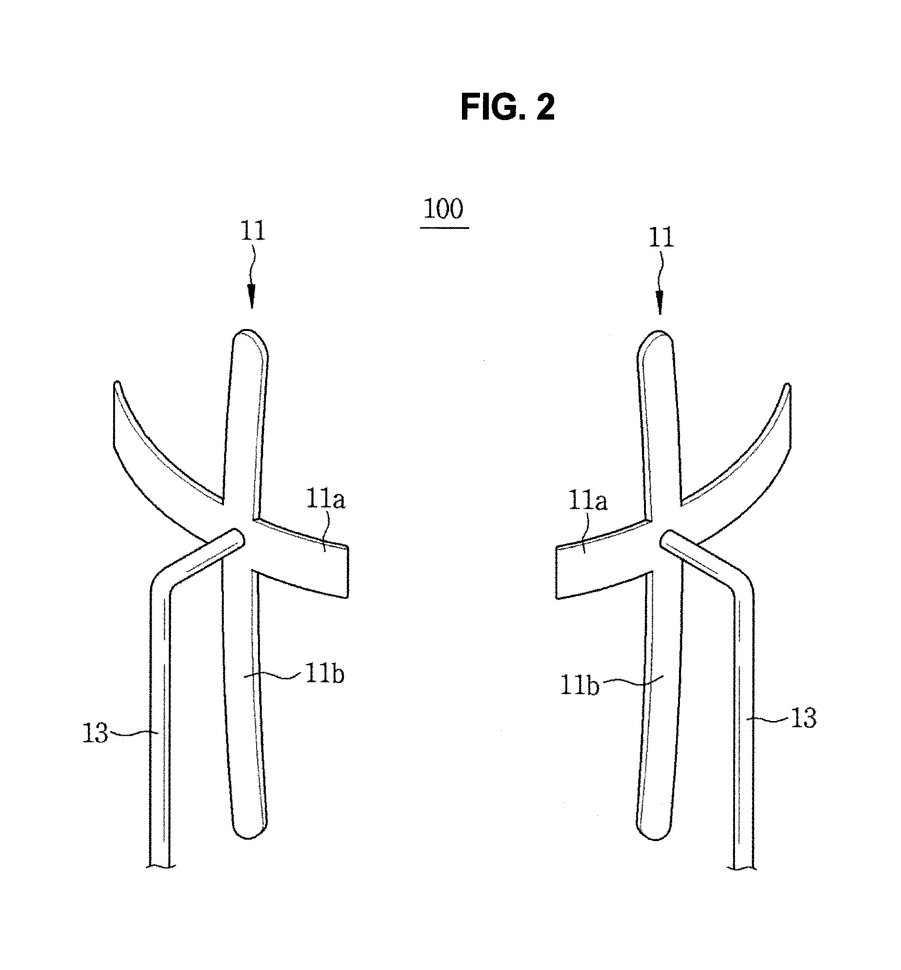
[0104]A head fixing apparatus according to embodiments to be described below (hereinafter, “head fixing apparatus”) serves to support the head of a subject during radiography, and includes an occipital region support for supporting the back of the head and a head holder provided to the occipital region support to prevent lateral movement of the head, wherein the head holder serves to support the temporal regions.

[0105]Specifically, the head holder is movably provided to the occipital region support and is tightened on the head to prevent lateral movement of the head. Thus, the head fixing apparatus may be used not only in radiography for acquisition of a head image but also in photography for acquisition of a facial image.

[0106]The head holder includes: a left support movably provided to the occipital region support to press a left side of the head; and a right support movably provided to the occipital region support to press a right side of the head. In other words, the head holder...


PUM


Abstract

Disclosed are a head fixing device, which is applied to an X-ray imaging system having an X-ray generator and an X-ray detector for photographing an image of a head and supports a head of an examinee such that the head of the examinee is maintained at a photographing position in an aligned state, and an X-ray imaging system using the same. Here, the head fixing device supports the occipital region and the temporal region of the examinee, thereby aligning the head of the examinee in the photographing position and maintaining the position of the head. The head fixing device comprises: an occipital region support (occipital support portion) for supporting the back of the head to block movement toward the rear side of the head, and a temporal region support (temporal support portion) for blocking left/right movement of the head, wherein the temporal region support may be provided in the occipital region support or may be separated from the occipital region support. In general radiographic inspection and dental radiographic inspection, the present invention can effectively fix the position of the head and ensure a stable position, thereby minimizing the probability of or preventing the generation of an error during radiography and obtaining an image having a high diagnostic value.

Description

TECHNICAL FIELD[0001]The present invention relates to a head positioning apparatus, i.e. a head fixing apparatus for holding the head of a subject in an imaging position during radiography, and more particularly, to a head fixing apparatus which allows the head to be properly positioned and stably held in place without causing deformation of facial soft tissues, and an x-ray imaging system including the same.BACKGROUND ART[0002]Photography using radiation, particularly, x-rays, is an important technique used to obtain internal images of a human body in medicine. An x-ray imaging apparatus refers to an imaging apparatus using x-rays.[0003]Such a typical x-ray imaging apparatus using x-rays is used in various forms to photograph all or some of a human body, i.e. to obtain images related to conditions of internal structures of the human body such as internal organs or bones, a structure of teeth, a skull, and the like.[0004]A typical x-ray imaging apparatus includes an x-ray generator ...

Claims


Application Information

Patent Timeline
IPC IPC(8): A61B90/14A61B6/00
CPCA61B6/501A61B90/14A61B6/0478A61B6/14A61B6/04
Inventor KIM, SEONG-HUNAHN, HYO-WONCHOI, YONG-SEOK
Owner UNIV IND COOP GRP OF KYUNG HEE UNIV
Why Eureka
Try Eureka