Device and method for monitoring internal organs

a technology for internal organs and devices, applied in the field of minimally invasive diagnosis of conditions within the body of an animal, can solve the problems of inability to present a reliable dynamic picture of the functional characteristics of the organ, inability to carry out ambulatory testing, and inability to meet the needs of ambulatory testing, so as to minimize the risk of infection, reduce the cost, and minimize the effect of discomfort and/or side effects

Inactive Publication Date: 2016-07-21
UTI LLP
View PDF9 Cites 0 Cited by
  • Summary
  • Abstract
  • Description
  • Claims
  • Application Information

AI Technical Summary

Benefits of technology

[0011]Some aspects of the invention provide a device and a method for monitoring internal organs using minimally invasive technique. In particular, the invention provides a device and a method for using transluminal electrical signal measurement to monitor internal organs. Such a device and method offer inter alia minimal invasive monitoring of internal organs thereby significantly reducing, the cost and minimizing or preventing possible discomfort and / or side-effects observed by conventional devices and methods.

Problems solved by technology

Impedance-based measurements of internal organs have been suggested in the prior art, but as a rule, they are not transluminal and transverse to a particular organ or interest, and because of that, they cannot present a reliable dynamic picture of the functional characteristics of the organ.
Scintigraphy is presently the most accurate and commonly used test, however it is expensive, time consuming, involves radiation, and is not fit for ambulatory testing.
It is particularly limited and affected by body position.
Many other tests are limited by cost or low accuracy, and do not offer a means to utilize transluminal transverse impedance measurements to assess gastric function.
However, this technology does not offer means to utilize transluminal, transverse impedance measurements to assess liver stiffness.
They utilize a non-transverse shear wave generator and receiver to assess liver stiffness, however the setups can be challenged in their ability to couple enough energy through the skin and subcutaneous fat to reach organs such as the liver, especially in obese patients.
This technology does not offer a means to utilize transluminal, transverse impedance measurements to assess liver stiffness.

Method used


Image

  • Device and method for monitoring internal organs
  • Device and method for monitoring internal organs
  • Device and method for monitoring internal organs

Examples


examples

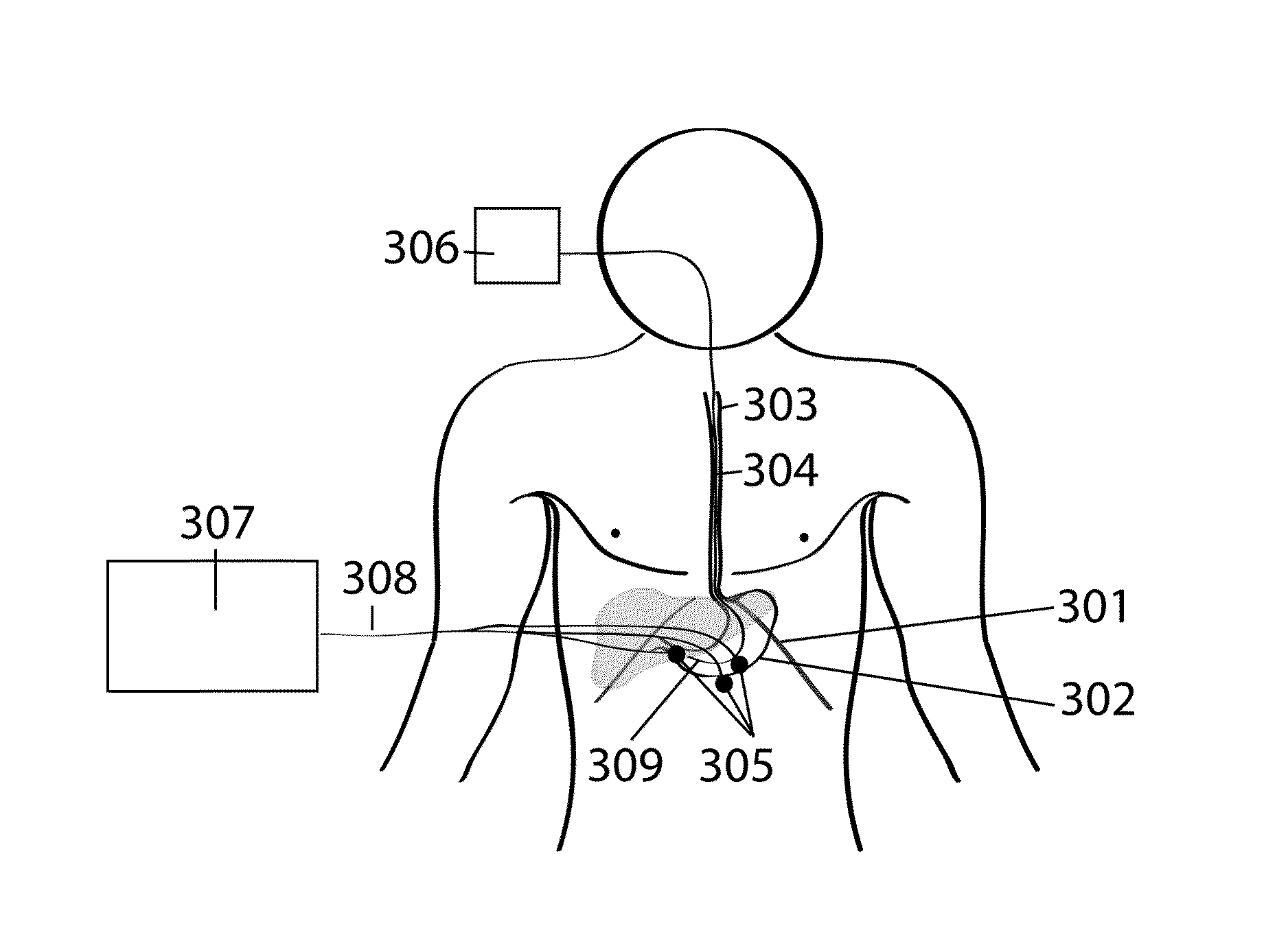
[0062]Materials and Methods: TIIM Capsule Design: For the present study the TIIM transducer was implemented as a gastric retentive pill, although it could also be positioned on the tip of a transnasal or transoral catheter. Each custom-designed TIIM transducer contained a miniature, battery-supplied, 50 kHz, 1.5 V oscillator (Linear Technology, Milpitas, Calif., USA). The length of the transducer was 18 mm with a diameter of 11 mm. It was embedded in dry, biocompatible super-absorbent polymer granules contained in a nonwoven permeable polyvinyl alcohol mesh (20-gsm) inside a size AAA DB capsule (Capsugel, Morristown, N.J., USA). The polymer granules swelled to 30-40 times their dry size in gastric liquid. The parameters of the expandable capsule were chosen to have minimal impact on the stomach and to maintain ease of swallowing, while still being able to ensure gastric retention, even during inter-digestive periods. The permeable gastric-retentive capsule design has self-disintegra...


PUM


Abstract

The present invention provides a method and an apparatus for minimally-invasive diagnosis of conditions within the body of an animal. In one particular embodiment, the present invention provides a method and an apparatus for utilizing intraluminally-generated signals to diagnose disorders or monitor the function of internal organ of an animal by assessing the signal transcutaneously. The signals can be electrical, magnetic, electromagnetic, acoustic, ultrasonic, optical, etc.

Description

CROSS-REFERENCE TO RELATED APPLICATIONS[0001]This application claims the priority benefit of U.S. Provisional Application No. 61 / 959,670 filed Aug. 30, 2013, which is incorporated herein by reference in its entirety.FIELD OF THE INVENTION[0002]The present invention relates generally to the minimally-invasive diagnosis of conditions within the body of an animal. In one particular embodiment, the present invention relates to generating a signal within the interior of the animal's body and assessing the signal transcutaneously to diagnose monitor or diagnose an internal organ of the animal. The signal can be electrical, magnetic, electromagnetic, acoustic, ultrasonic, optical, etc.BACKGROUND OF THE INVENTION[0003]Impedance-based measurements of internal organs have been suggested in the prior art, but as a rule, they are not transluminal and transverse to a particular organ or interest, and because of that, they cannot present a reliable dynamic picture of the functional characteristic...

Claims


Application Information

Patent Timeline
Patent Type & Authority Applications(United States)
IPC IPC(8): A61B5/00
CPCA61B5/0031A61B5/6852A61B5/6861A61B5/0028A61B5/0538A61B5/4233A61B5/4238A61B5/4244A61B5/4255
Inventor POSCENTE, MICHAEL D.YADID-PECHT, ORLYANDREWS, CHRISTOPHER N.MINTCHEV, MARTIN P.
Owner UTI LLP
Why Eureka
Try Eureka