Method and apparatus for adjusting corneal curvature through digital corneal crosslinking
a crosslinking and digital technology, applied in the field of adjusting corneal curvature, can solve the problems of invasive process, loss of ability to focus images on the retina, refractive error, or ametropia, etc., and achieve the effect of accurately and efficiently adjusting the corneal curvature and minimizing invasiveness
- Summary
- Abstract
- Description
- Claims
- Application Information
AI Technical Summary
Benefits of technology
Problems solved by technology
Method used
Image
Examples
example 1
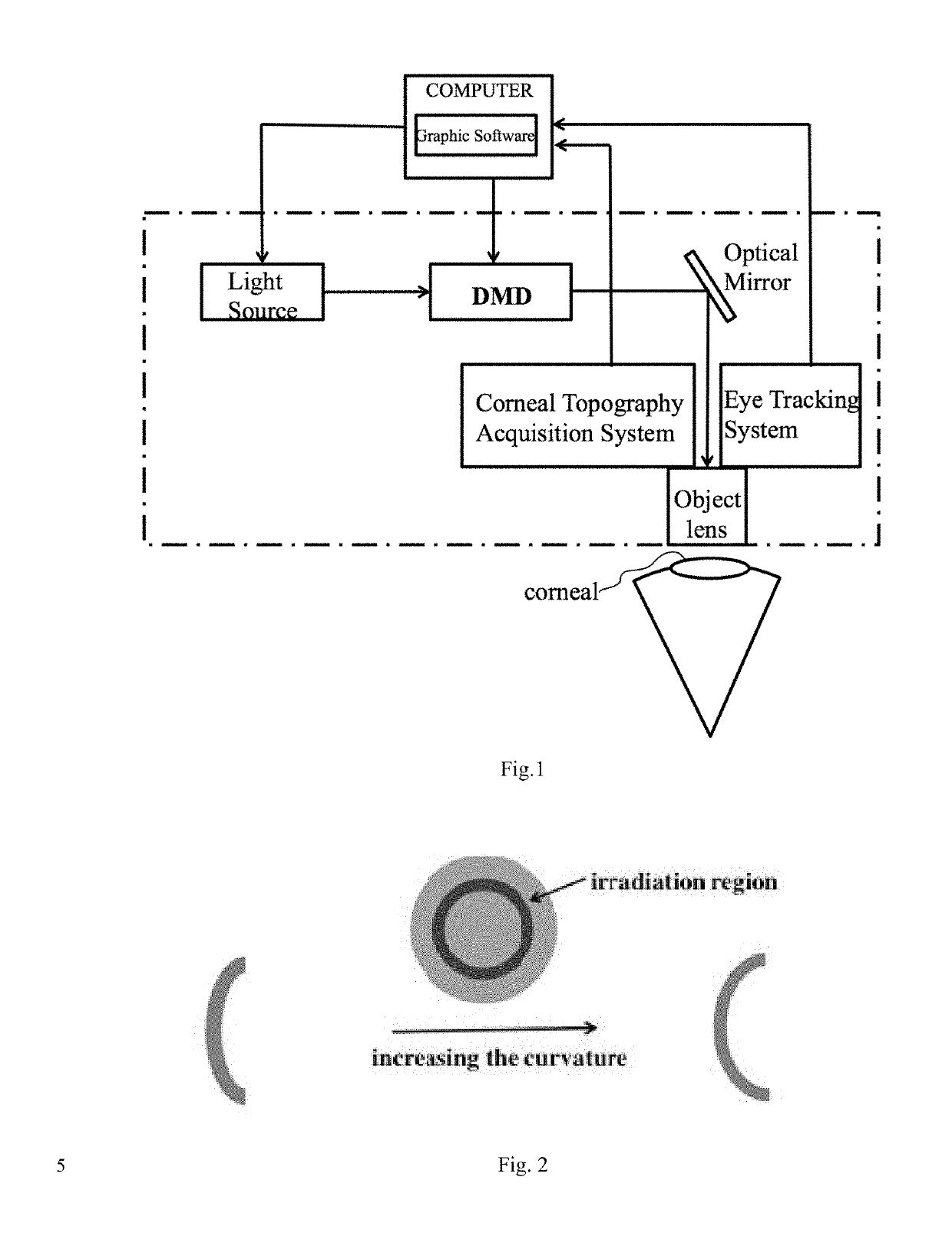
[0041]The epithelial tissue of an area of 7 mm diameter in the middle of the cornea was removed after superficial anesthesia. A dextran solution of 200 g / L with 1 g / L riboflavin was dropped to corneal surface in batches. The location of the riboflavin diffused into the corneal surface was observed by cobalt blue light of a slit lamp. Digital corneal crosslinking device was used to selectively irradiate the local area of the cornea. The light's wavelength is 365 nm, the irradiation area is shown in FIG. 2, the light power is 1.2 mW / cm2, irradiation time is 5 minutes.
[0042]During irradiation process, a photoinitiator solution and superficial anesthesia were used to wash the corneal surface in phases. Antibiotic eye formulations were applied to the eye and corneal contact lens were used until the corneal epithelium was healed. As shown in FIG. 2, the corneal surface curvature increased.
example 2
[0043]The epithelial tissue of an area of 7 mm diameter in the middle of the cornea was removed after superficial anesthesia. A dextran solution of 200 g / L with 1 g / L riboflavin sodium phosphate was dropped to corneal surface in batches. The location of the riboflavin diffused into the corneal surface was observed by cobalt blue light of a slit lamp. Digital corneal crosslinking device was used to selectively irradiate the local area of the cornea. The light's wavelength is 365 nm, the irradiation area is the same like that in example 1, the light power is 1.2 mW / cm2, irradiation time is 5 minutes.
[0044]During irradiation process, a photoinitiator solution and superficial anesthesia were used to wash the corneal surface in phases. Antibiotic eye formulations were applied to the eye and corneal contact lens were used until the corneal epithelium was healed. The corneal surface curvature was the same as example 1.
example 3
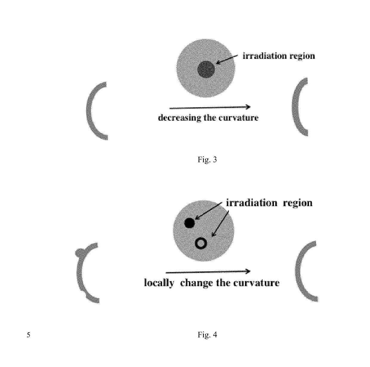
[0045]The epithelial tissue of an area of 5-9 mm diameter in the middle of the cornea was removed after superficial anesthesia. A dextran solution of 200 g / L with 1 g / L eosin Y was dropped to corneal surface in batches. The location of the eosin Y diffused into the corneal surface was observed by cobalt blue light of a slit lamp. Digital corneal crosslinking device was used to selectively irradiate the local area of the cornea. The light's wavelength is 550 nm, the irradiation area is shown in FIG. 3, the light power is 1.2 mW / cm2, irradiation time is 5 minutes.
[0046]During irradiation process, the eosin Y solution and superficial anesthesia were used to wash the corneal surface in phases. Antibiotic eye formulations were applied to the eye and corneal contact lens were used until the corneal epithelium was healed. As shown in FIG. 3, the corneal surface curvature decreased.
PUM
| Property | Measurement | Unit |
|---|---|---|
| wavelength | aaaaa | aaaaa |
| wavelength | aaaaa | aaaaa |
| time | aaaaa | aaaaa |
Abstract
Description
Claims
Application Information
 - R&D
- Intellectual Property
- Life Sciences
- Materials
- Tech Scout
- Unparalleled Data Quality
- Higher Quality Content
- 60% Fewer Hallucinations
Browse by: Latest US Patents, China's latest patents, Technical Efficacy Thesaurus, Application Domain, Technology Topic, Popular Technical Reports.
© 2025 PatSnap. All rights reserved.Legal|Privacy policy|Modern Slavery Act Transparency Statement|Sitemap|About US| Contact US: help@patsnap.com


