Imaging and measuring system of vocal cord vibration based on plane wave ultrasonography, and method thereof

a plane wave ultrasonography and vibration imaging technology, applied in the field of biomedicine information detection, can solve the problems of egg being unable to describe the vibration properties of a certain tissue region patient cannot pronounce in a natural voice, and all cannot provide vibration imaging of internal organizational structures below the surface of the vocal cord, etc., to improve the accuracy of the glottis closure quotient, and improve the accuracy of the effect of the glo

Inactive Publication Date: 2016-10-13
XI AN JIAOTONG UNIV
View PDF6 Cites 2 Cited by
  • Summary
  • Abstract
  • Description
  • Claims
  • Application Information

AI Technical Summary

Benefits of technology

The present invention solves the problem of motion blur in conventional ultrasonography technologies by using a plane wave sending method instead of a linear scanning method. This results in a laryngeal tissue structure image in an entire plane, which is suitable for researches of aperiodic irregular vibration of the vocal cords under a non-stable pronouncing condition. The feature points are available from the UGG, which is highly reliable during extraction. The present invention is non-invasive and ensures that the testee is able to naturally pronounce and dynamically pronounce. The plane wave imaging technologies and point synchronization technologies eliminate space asynchronism and time randomness caused by ultrasound during sampling the vocal cord vibration. Thus, the present invention provides the time-space synchronization of the vocal cord vibration detection.

Problems solved by technology

However, conventionally, researches of problems such as how the vocal cords in the body regulates biomechanical properties thereof to change a sound mode, and how lesions change the biomechanical properties of the vocal cords to caused pathological voice, are still in an infancy.
However, optical imaging technologies for larynx and vocal cords, comprising strobe dynamic laryngoscope and high-speed photography laryngoscope, are all not able to provide vibration imaging of internal organizational structures below surfaces of the vocal cords.
Furthermore, due to invasion of the endoscope of the optical device, a patient is not able to pronounce in a natural voice.
Therefore, the EGG is unable to describe quantify vibration properties of a certain tissue region of the vocal cords.
However, a conventional ultrasonography technique uses a line-by-line scan mode, which divides an image into a number of scan lines, while data of each scan line are obtained at different moments, which leads to certain time differences between different points of the image during sampling.
When it comes to the high-speed vibration of the vocal cords, the time differences cannot be ignored.
In this case, the image will be blurred because of the high-speed vibration of the vocal cords, resulting in uncertain measurement of vibration velocity and displacement of the vocal cords.
In addition, because of a low imaging frame rate (<1000 Hz) of the conventional ultrasonography technique, requirements of vocal cord vibration under unstable situations are unable to be satisfied.
A transmitting beam of the single array element ultrasound transducer has a strong directivity, which is not able to detect an overall structure and positions of the vocal cords.
Without image guide, the single array element ultrasound transducer is easy to lose information during detection.
However, a linear array transducer, which is capable of imaging of the vocal cord vibration within a range of an entire vocal cord length, has a limited application.
Since the UGG reflects phase information of the vocal cord vibration, asynchronous problem is unacceptable.
So, high-speed imaging of the vocal cord vibration with time and space synchronization, and quantitatively extracting information of biomechanical parameters as well as certain phase changes are still major problems in the field.

Method used


Image

  • Imaging and measuring system of vocal cord vibration based on plane wave ultrasonography, and method thereof
  • Imaging and measuring system of vocal cord vibration based on plane wave ultrasonography, and method thereof
  • Imaging and measuring system of vocal cord vibration based on plane wave ultrasonography, and method thereof

Examples


Embodiment Construction

[0055]Referring to the drawings and a preferred embodiment, the present invention is further illustrated.

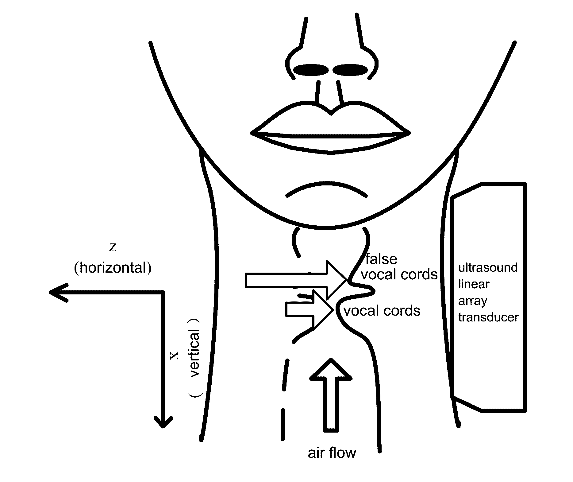
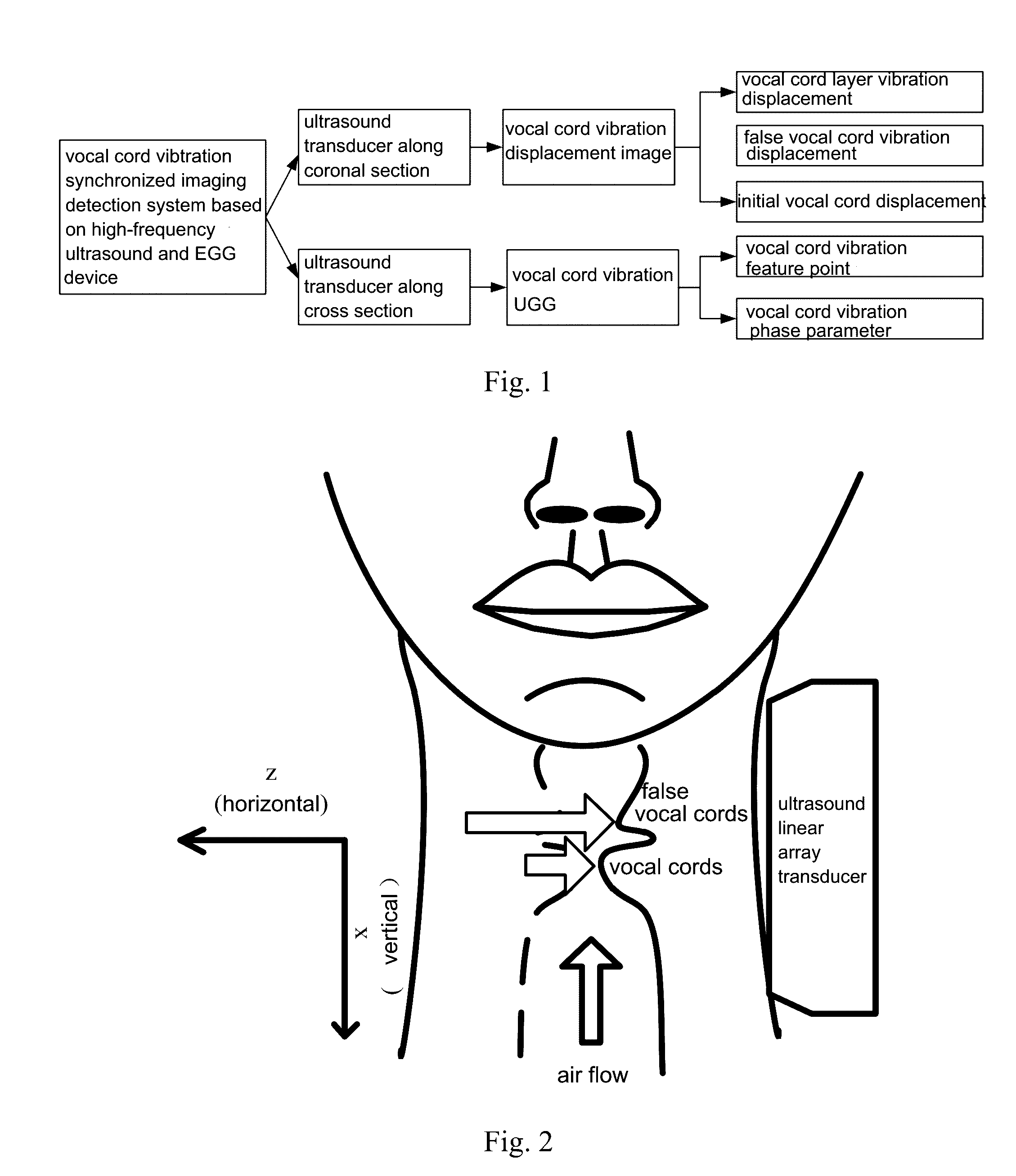
[0056]Referring to FIGS. 1-7, an imaging and measuring system of vocal cord vibration based on plane wave ultrasonography, comprising: a digital ultrasonography system, a data acquisition unit, and a computer.

[0057]The digital ultrasonography system comprises an ultrasound linear array transducer and a host; wherein the ultrasound linear array transducer is controlled by the host for sending an ultrasound plane wave and receiving an echo; the echo is sent back to the host; wherein the host sends back the echo to the data acquisition unit; wherein the data acquisition unit converts a received echo signal into a digital signal and then sends to the computer; wherein the computer provides beam formation of echo data, radio frequency signal envelope detection, and dynamic range compression of the digital signal received, for obtaining a laryngeal tissue structure image.

1) Overall Pro...


PUM


Abstract

An imaging and measuring system of vocal cord vibration based on plane wave ultrasonography, includes: a digital ultrasonography system, a data acquisition unit, and a computer; wherein the digital ultrasonography system comprises an ultrasound linear array transducer and a host; wherein the ultrasound linear array transducer is controlled by the host for sending an ultrasound plane wave and receiving echo; the echo is sent back to data acquisition unit; wherein the data acquisition unit converts the echo into digital signal and then sends it to the computer; wherein the computer provides beamforming, envelope detection, and dynamic range compression of the digital signal received, for obtaining a laryngeal tissue structure image. The present invention provides high-speed imaging of the vocal cord vibration with temproal and spatial synchronization, and quantitatively extracting information of biomechanical parameters as well as vibrational phase changes.

Description

CROSS REFERENCE OF RELATED APPLICATION[0001]This is a U.S. National Stage under 35 U.S.C 371 of the International Application PCT / CN2014 / 094449, filed Dec. 19, 2014, which claims priority under 35 U.S.C. 119(a-d) to CN 201410605785.5, filed Oct. 30, 2014.BACKGROUND OF THE PRESENT INVENTION[0002]1. Field of Invention[0003]The present invention relates to a field of biomedicine information detection, and more particularly to high-speed vibration imaging which provides time-space synchronization of vocal cords, as well as a system for quantitatively extracting time-space vibration properties of the vocal cords and a method thereof.[0004]2. Description of Related Arts[0005]High-speed, complex and multi-dimensional vibration of human vocal cords generates a voice source. The vocal cords are small organs with highest vibration speeds of a human body, but are also most vulnerable organs. However, conventionally, researches of problems such as how the vocal cords in the body regulates biome...

Claims


Application Information

Patent Timeline
IPC IPC(8): A61B8/08A61B8/00A61B8/14
CPCA61B8/08A61B8/5223A61B8/4483A61B8/14A61B8/0858A61B8/5284
Inventor WAN, MINGXITANG, SHANSHANJING, BOWENWANG, SUPIN
Owner XI AN JIAOTONG UNIV
Why Eureka
Try Eureka