Characterization of respiratory motion in the abdomen using a 4d MRI technique with 3D radial sampling and respiratory self-gating

a motion and abdominal organ technology, applied in the field of imaging methods and systems, can solve the problems of poor treatment outcomes, under-dosage in tumors and/or over-dosage in healthy tissues, and respiratory motion in abdominal organs poses significant challenges to accurate determination of treatment margins for target tumors

Active Publication Date: 2016-11-10
CEDARS SINAI MEDICAL CENT
View PDF7 Cites 30 Cited by
  • Summary
  • Abstract
  • Description
  • Claims
  • Application Information

AI Technical Summary

Problems solved by technology

In free-breathing radiotherapy treatment planning, respiration-induced motion in abdominal organs poses significant challenges to accurate determination of treatment margins for target tumors.
Inaccurate margin prescription may potentially result in under-dosage in tumors and / or over-dosage in healthy tissues, which may lead to poor treatment outcomes.
However, 4D-CT has a number of limitations including: 1) high dose of ionizing radiation caused by repetitive irradiation at the same location necessary to image a complete respiratory cycle; 2) suboptimal tumor delineation due to poor soft tissue contrast; 3) and most importantly, susceptibility to manifest motion artifacts (i.e. resorting artifacts) caused by breathing pattern variation between individual 2D slice acquisitions.
However, this approach may not fully characterize the 3D motion of the entire tumor, making it difficult to accurately calculate the ITV for irregularly-shaped or deformable tumors.
In addition, healthy vulnerable tissues surrounding the tumor may not be fully defined.
The real-time 3D techniques are limited by the achievable spatiotemporal resolution, which may cause blurring of the fine or fast-moving structures.
These 2D approaches have relatively low slice resolution of 5-10 mm, which may limit the accurate depiction of respiratory motion in the through-slice direction.
More importantly, irregular breathing, commonly seen in patients, could potentially elicit unpredictable and prohibitively long scan times for the prospectively gated techniques and induce severe resorting artifacts, as in 4D-CT, for the retrospectively gated techniques.
This technique, however, is inherently limited in the respiratory motion-sampling rate (˜250 ms), which could potentially result in errors in the respiratory curve during deep or fast breathing and hence large intra-phase motion variability.

Method used


Image

  • Characterization of respiratory motion in the abdomen using a 4d MRI technique with 3D radial sampling and respiratory self-gating
  • Characterization of respiratory motion in the abdomen using a 4d MRI technique with 3D radial sampling and respiratory self-gating
  • Characterization of respiratory motion in the abdomen using a 4d MRI technique with 3D radial sampling and respiratory self-gating

Examples


example 1

Methods

Sequence Design

[0037]A spoiled gradient recalled echo (GRE) sequence with 3D radial-sampling k-space trajectory and one-dimensional (1D) projection-based SG was implemented at 3.0 T for image acquisition (see Pang J, Sharif B, Fan Z, Bi X, Arsanjani R, Berman D S, Li D. ECG and navigator-free four-dimensional whole-heart coronary MRA for simultaneous visualization of cardiac anatomy and function. Magn Reson Med 2014, which is incorporated herein by reference in its entirety as though fully set forth).

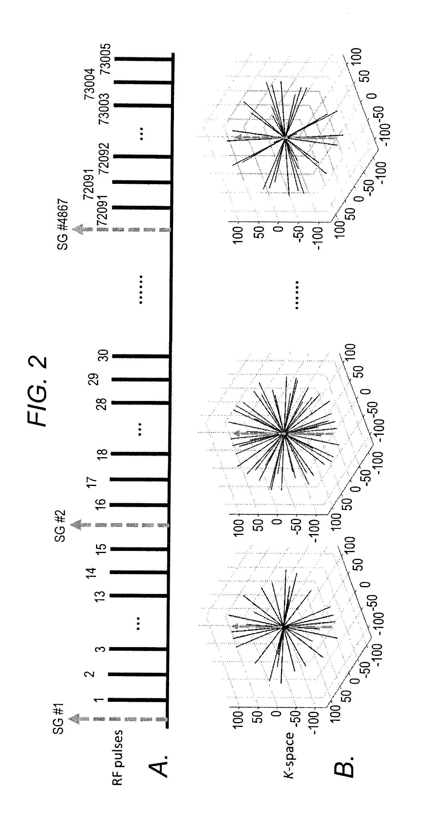
[0038]In an attempt to preserve the sampling pattern stability required for arbitrary retrospective data sorting, the 3D radial k-space was filled using the 2D golden means ordering (see Chan RW, Ramsay E A, Cunningham C H, Plewes D B. Temporal stability of adaptive 3D radial MRI using multidimensional golden means. Magnetic Resonance in Medicine 2009; 61:354-363, which is incorporated herein by reference in its entirety as though fully set forth), a generalization of the origina...

example 2

Experiments

[0047]The feasibility of the technique was demonstrated with motion phantom studies and in-vivo liver imaging studies. All scans were performed on a clinical 3.0 T MRI system (MAGNETOM Verio, Siemens Healthcare, Erlangen, Germany) equipped with a 32-channel receiver coil (Invivo, Gainesville, Fla., USA).

Study Design

[0048]Schematics of the motion phantom are shown in FIG. 4a. A commercial Dynamic Breathing Phantom (RSD™, Long Beach, Calif., USA) system placed outside the scanner room was used to produce simulation signals that mimic the human respiratory motion. Through an air pump and tube, the signals were used to drive a plastic box (35×40×63 mm3) fully filled with gadolinium-doped water and sealed with silicone sealant. Five PinPoint® fiducial markers (Beekley Medical™, Bristol, Conn., USA) were placed on each face of the box. The box was then used as an imaging target, where reciprocating motion was executed along the z-axis of the magnet at a frequency of 18 cycles / m...

example 3

Results

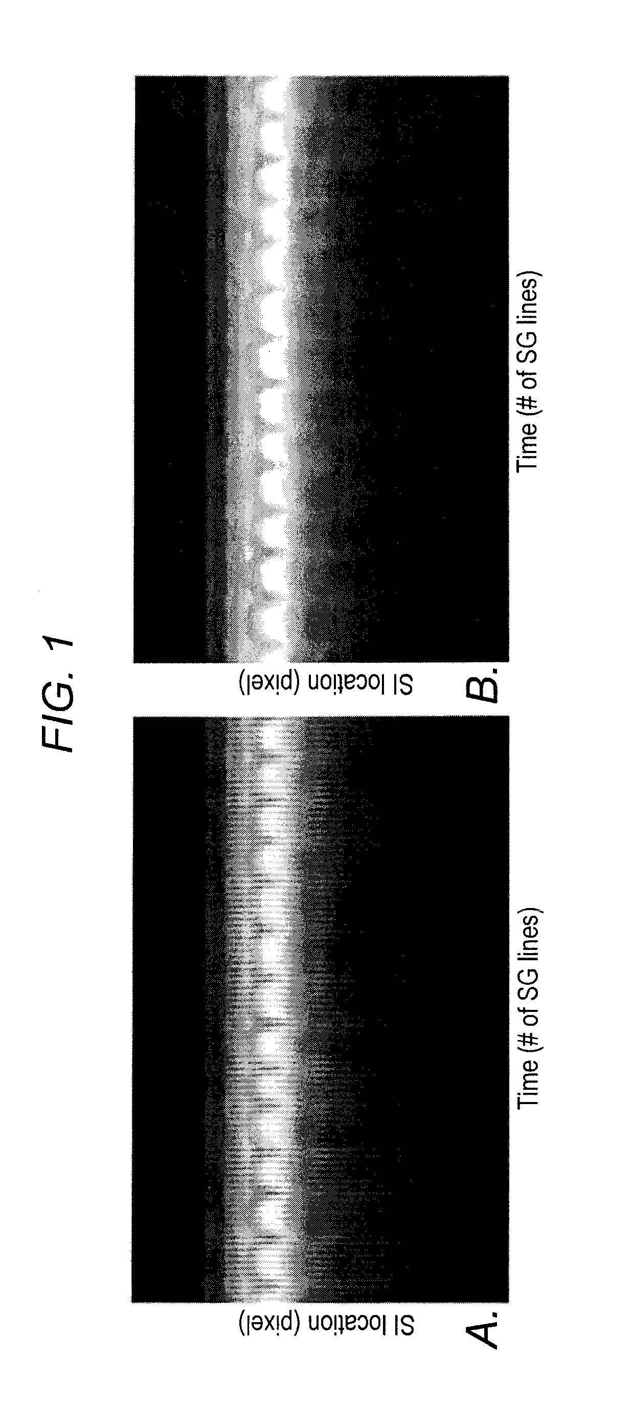
[0058]The SI motion of the imaging targets was well appreciated on the SG-derived respiratory curves. As expected, the motion phantom showed a strictly sinusoidal respiratory pattern and the human subjects, as illustrated in FIG. 3a (a healthy subject) and FIG. 3b (a patient), showed variations in breathing amplitude and frequency, with the patient showing more irregularity than the healthy volunteer. The retrospective data sorting strategy described in the examples above detected the breathing cycles with abnormal respiratory durations (rectangles on the zoom-in respiratory curve in FIG. 3) or expiratory peaks (vertical ovals) and also identified the outlier with large respiratory phase drift (horizontal oval). As a result, 5-16% of the total k-space data were discarded, with approximately 6000-7000 imaging lines available in each respiratory phase for image reconstruction.

[0059]The method in the non-limiting example set forth above offered respiratory phase-resolved volumet...


PUM


Abstract

In some embodiments, the present application discloses utilizing a continuous spoiled gradient echo sequence with 3D radial trajectory and 1D self-gating for respiratory motion detection. In certain embodiments, data acquired are retrospectively sorted into different respiratory phases based on their temporal locations within a respiratory cycle, and each phase is reconstructed via a self-calibrating CG-SENSE program.

Description

FIELD OF THE INVENTION[0001]The present invention generally relates to imaging methods and systems.BACKGROUND[0002]The following description includes information that may be useful in understanding the present invention. It is not an admission that any of the information provided herein is prior art or relevant to the presently claimed invention.[0003]In free-breathing radiotherapy treatment planning, respiration-induced motion in abdominal organs poses significant challenges to accurate determination of treatment margins for target tumors. Inaccurate margin prescription may potentially result in under-dosage in tumors and / or over-dosage in healthy tissues, which may lead to poor treatment outcomes. As one of the important steps in the determination of treatment margins, the internal target volume (ITV), derived from the union of three-dimensional (3D) volumes of the tumor at all respiratory phases is needed to account for the variation of the tumor in position, shape, and size due ...

Claims


Application Information

Patent Timeline
Patent Type & Authority Applications(United States)
IPC IPC(8): A61B8/08A61B8/00
CPCA61B8/085A61B8/483A61B8/54A61B8/5215A61B8/5207A61B5/055A61B5/7207A61B5/113A61B5/7257G01R33/4826G01R33/5611G01R33/56509G01R33/5676
Inventor FAN, ZHAOYANGPANG, JIANINGDENG, ZIXINLI, DEBIAO
Owner CEDARS SINAI MEDICAL CENT
Why Eureka
Try Eureka