Detecting a Therapeutic Cell

Active Publication Date: 2017-03-02
UCL BUSINESS PLC
View PDF0 Cites 6 Cited by
  • Summary
  • Abstract
  • Description
  • Claims
  • Application Information

AI Technical Summary

Benefits of technology

0042]The present invention therefore enables therapeutic cells in the CNS to be detected and/or quantified. Examples of such therapeutic cells include therapeutic cells which are administered to a subject as a medicament, and hos

Problems solved by technology

Nevertheless, the fate of the therapeutic T cells in clinical studies remains unclear, particularly in central nervous system (CNS) where the engraftment of the T-cells is appreciably more challenging to measure.
Despite the continued advancements in monitoring reporter gene expressing T-cells in the peripheral nervous system there is no translatable or clinical standard for tracking cells in the CNS, in particular in the brain.
This

Method used


Image

  • Detecting a Therapeutic Cell
  • Detecting a Therapeutic Cell
  • Detecting a Therapeutic Cell

Examples


example 1

Functional Expression and Binding of Dopamine Active Transporter (DAT) in T-Cells

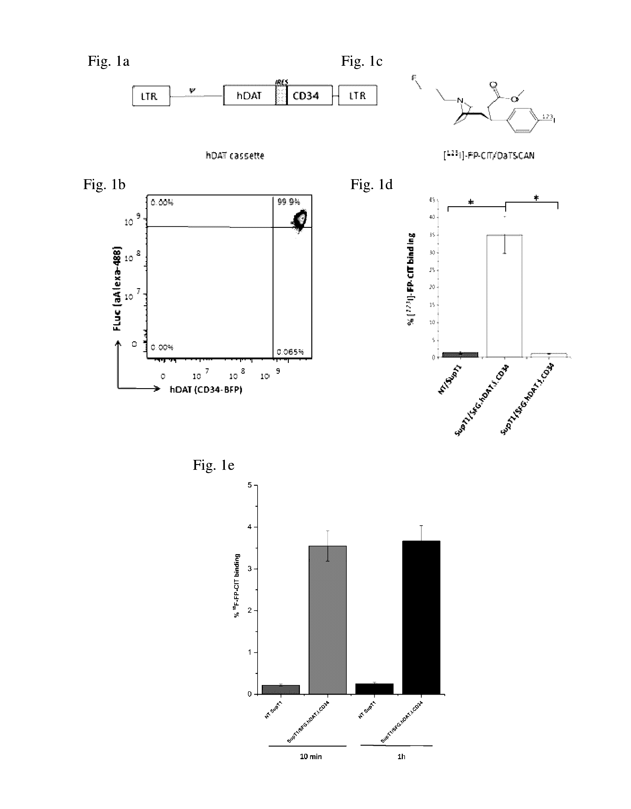
[0216]To determine the potential for utilising a human DAT (hDAT) reporter to image T-cells using [123I]-FP-CIT, a retroviral vector (SFG.hDAT.I.CD34) encoding both hDAT and CD34 genes was constructed (FIG. 1a) and used to transduce SupT1 cell line. The SupT1 cell line is a CD4+ human T-cell line with tumorigenic properties and stable transduction has been previously demonstrated with no significant reduction in the expression of introduced genes8. Flow analysis of the SupT1 / SFG.hDAT.I.CD34 cells indicated a transduction efficiency of 20-25% and cells were subsequently selected by CD34 expression to 99% purity using FACS (FIG. 1b).

[0217]Binding of [123I]-FP-CIT (FIG. 1c) to hDAT reporter cells was demonstrated by co-incubation of tracer with SupT1 / SFG.hDAT.I.CD34 cells and compared against control non-transduced (NT) SupT1 cells. The percentage [123I]-FP-CIT binding was 25 fold greater (FIG. 1d) in SupT...

example 2

T Cells Expressing hDAT Retain Proliferative and IFN-γ Release Ability

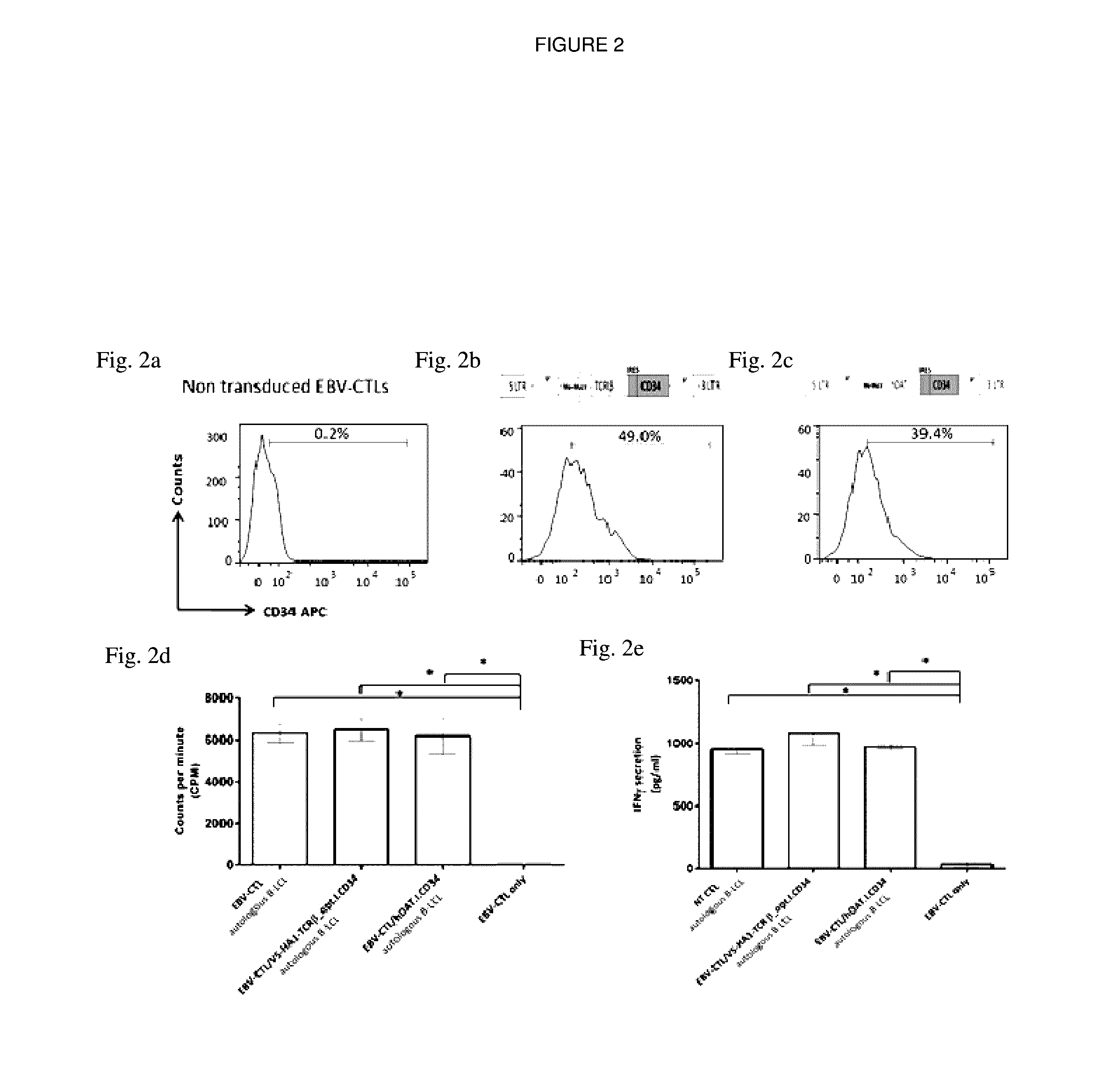
[0218]Primary T cells, EBV-specific CTLs were engineered with hDAT to study any effect the reporter may have on the proliferation and cytokine release. EBV-CTLs were transduced with hDAT (EBV-CTL / SFG.hDAT.I.CD34) or a control vector encoding a HA1 T cell receptor (EBV-CTL / SFG.V5-HA1-TCRβ_opt.I.CD34) and the proliferative and IFN-γ release ability was compared to non-transduced EBV-CTLs (NT / EBV CTLs). The transduction efficiency was 39.4% in EBV-CTL / SFG.hDAT.I.CD34 (FIG. 2b), 49.0% in EBV-CTL / SFG.V5-HA1-TCRβ_opt.I.CD34 (FIG. 2c) in comparison to NT / EBV CTLs (FIG. 2a). There was no significant (p>0.05) difference in the functional capabilities between NT / EBV CTLs, EBV-CTL / SFG.hDAT.I.CD34 and EBV-CTL / SFG.V5-HA1-TCRβ_opt.I.CD34 cells upon stimulation with B-LCLs indicating EBV-CTL / SFG.hDAT.I.CD34 cells were able to retain their proliferative and IFN-γ release ability at levels comparable to control cells.

example 3

In Vivo Evaluation of the hDAT / [123I]-FP-CIT Reporter System

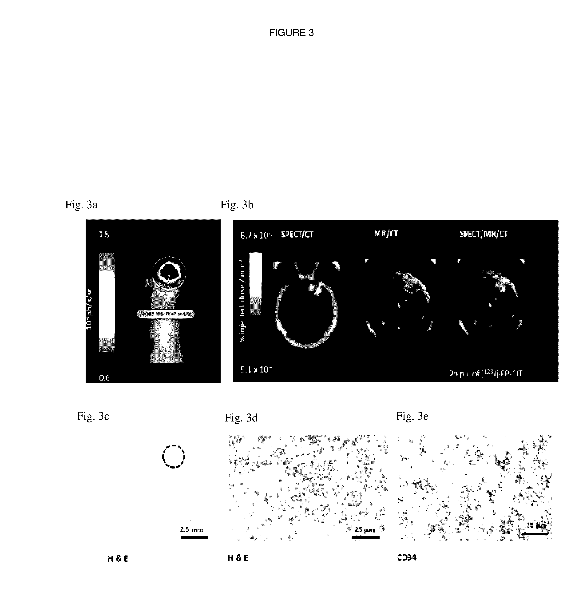
[0219]Immunocompromised mice (n=4) were stereotactically injected with SupT1 / SFG.hDAT.I.CD34 cells co-transduced with FLuc above the right basal ganglia. Sequential bioluminescence, MRI and SPECT images were acquired at day 25 post cell inoculation. Bioluminescence and MR images were acquired prior to [123I]-FP-CIT imaging. Cell viability was confirmed using bioluminescence (FIG. 3a) and xenograft volumes (22.86±4.96 mm3) were measured by manual drawing of ROIs on T2-weighted MR images (FIG. 3b).

[0220]The co-registered SPECT-MR-CT images displayed good co-localisation of the MR contrast and SPECT signal at the site of injection (FIG. 3b). Dynamic brain SPECT images were acquired at 15 min, 2 h, 4 h and 6 h post radiotracer injection. hDAT-positive xenografts were visualised as early as 15 mins and up to 6 h post [123I]-FP-CIT injection (FIG. 4a). A SPECT signal was non-detectable in control animals intracranially injected w...


PUM

PropertyMeasurementUnit
Fractionaaaaaaaaaa
Timeaaaaaaaaaa
Compositionaaaaaaaaaa

Abstract

The present invention relates to a method of detecting a therapeutic cell expressing a dopamine transporter (DAT) at a central nervous system (CNS) site in a subject, which comprises the administration of a DAT tracer to the subject, wherein the presence of a therapeutic cell which expresses the DAT is determined.

Description

FIELD OF THE INVENTION[0001]The present invention relates to methods and reagents which can be used to enable the detection of a therapeutic cell in the central nervous system of a subject.BACKGROUND TO THE INVENTION[0002]Malignant brain tumours are rapidly progressive and resistant to most treatments. In the last eighty years, long-term survival has improved from just six to eighteen months even with state-of-the-art standard of care (surgery, chemotherapy, and radiotherapy). Neurodegenerative disease such as Parkinson's Disease and Alzheimer's Disease also represent unmet clinical needs.[0003]With the promise of new cell based therapies, there is an urgent need for non-invasive, readily available and translatable methods of detecting and quantifying cells in vivo. The development of this imaging technology will enable visualization of biological processes where the fate, localization and long term viability of therapeutic cells can be monitored, which will greatly increase the val...

Claims


Application Information

Patent Timeline
IPC IPC(8): A61K51/04C07K16/28A61K39/395C07K14/47A61K38/17
CPCA61K51/0455C07K14/4702A61K38/1709A61K39/3955C07K16/2818C07K16/2809C07K16/2896C07K2317/622C07K2319/74C07K2319/02A61K39/0011A61K2039/5156A61K2039/5158C07K14/705C07K14/7051C07K2319/03
Inventor PULÉ, MARTINBADAR, ADAMKIRU, LOUISELYTHGOE, MARKPETERS, ADRIEN
Owner UCL BUSINESS PLC
Why Eureka
Try Eureka