Magnetic resonance imaging system with integrated photon detector ring

Active Publication Date: 2017-05-18
KONINKLJIJKE PHILIPS NV
View PDF9 Cites 2 Cited by
  • Summary
  • Abstract
  • Description
  • Claims
  • Application Information

AI Technical Summary

Benefits of technology

The invention provides a magnetic resonance imaging gradient coil assembly and a magnetic resonance imaging system that can incorporate a positron emission tomography or SPECT system without decreasing the useable bore of the magnet. This may save space within the magnet and provide more space for patients or subjects using the magnetic resonance imaging system.

Problems solved by technology

PET and SPECT have the disadvantage that they do not provide details about the subjects internal anatomy and the response of the body to the radiopharmaceutical is relatively slow.

Method used


Image

  • Magnetic resonance imaging system with integrated photon detector ring
  • Magnetic resonance imaging system with integrated photon detector ring
  • Magnetic resonance imaging system with integrated photon detector ring

Examples


Embodiment Construction

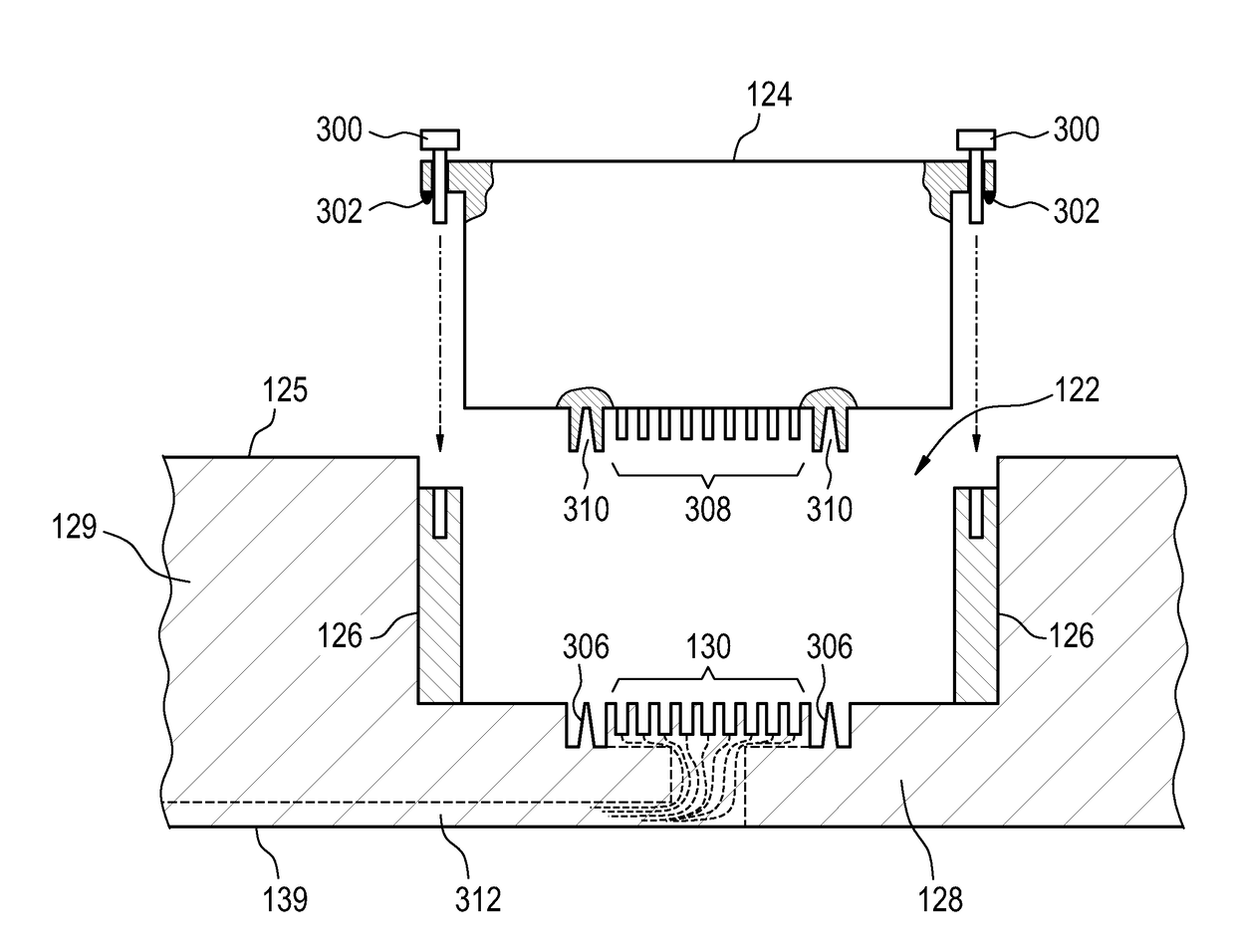
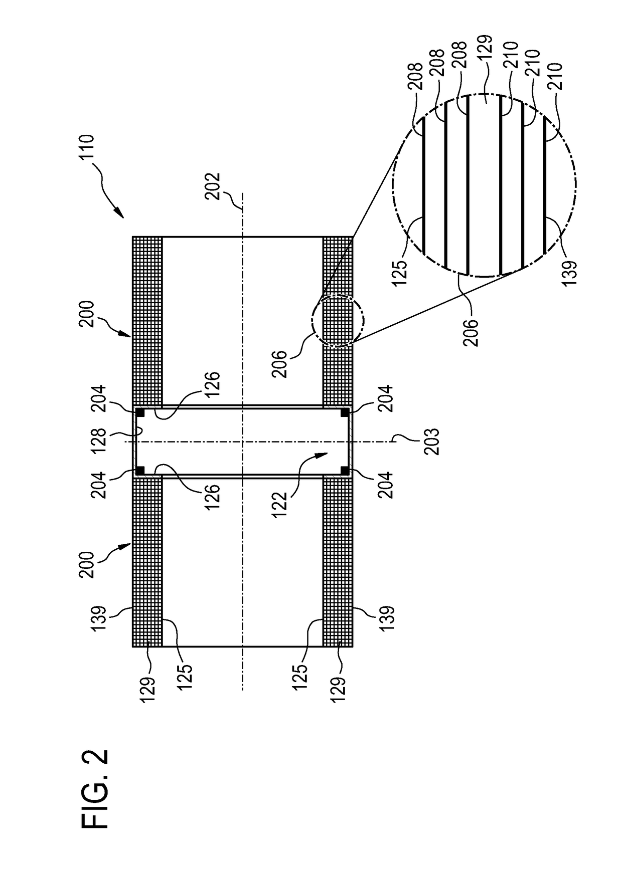
[0059]Like numbered elements in these figures are either equivalent elements or perform the same function. Elements which have been discussed previously will not necessarily be discussed in later figures if the function is equivalent.

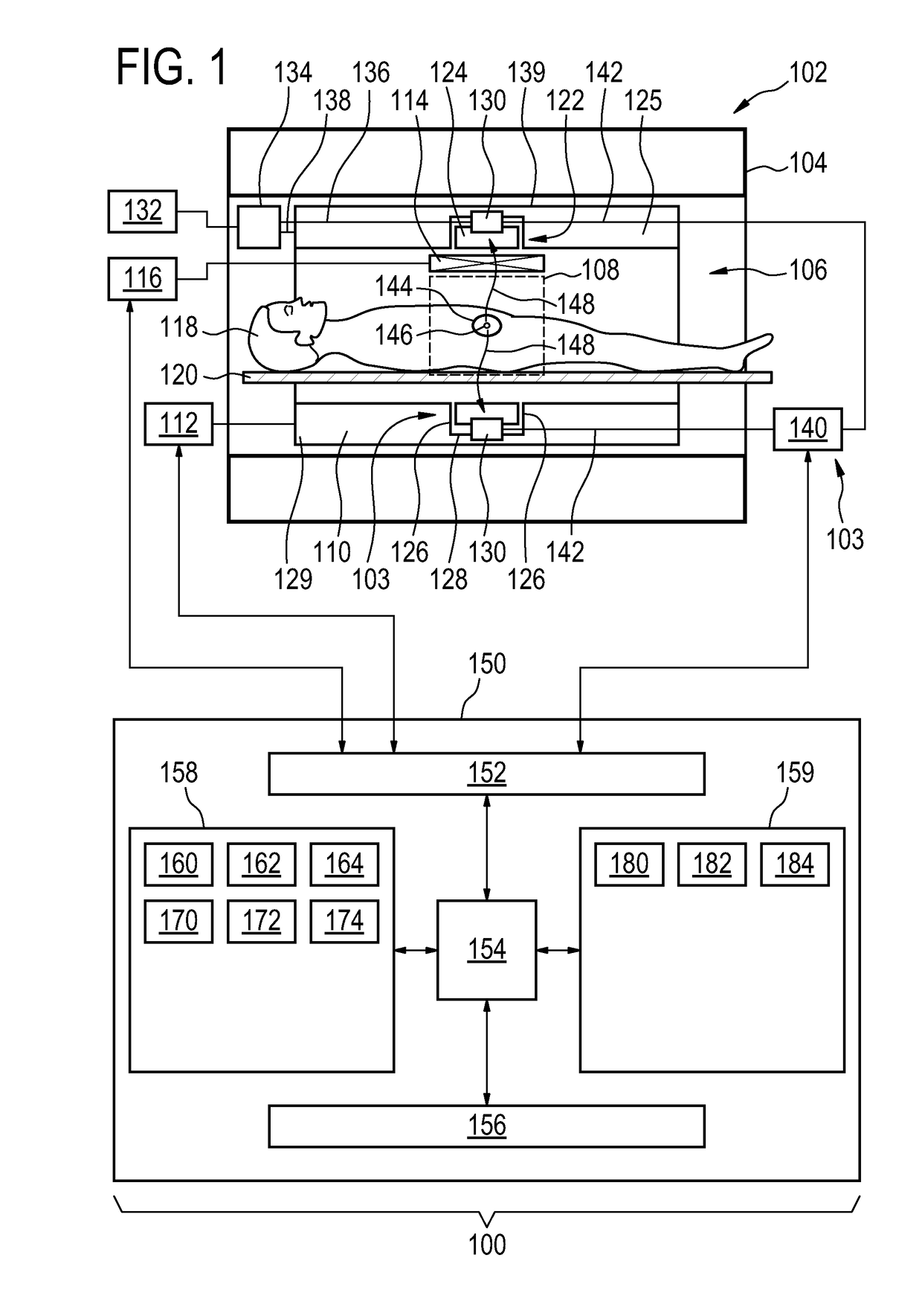
[0060]FIG. 1 illustrates an example of a medical instrument 100. The medical instrument 100 comprises magnetic resonance imaging system 102 with a magnet 104. The magnet 104 is a superconducting cylindrical type magnet 104 with a bore 106 through it. The use of different types of magnets is also possible for instance it is also possible to use both a split cylindrical magnet and a so called open magnet. A split cylindrical magnet is similar to a standard cylindrical magnet, except that the cryostat has been split into two sections to allow access to the iso-plane of the magnet, such magnets may for instance be used in conjunction with charged particle beam therapy. An open magnet has two magnet sections, one above the other with a space in-between that ...


PUM


Abstract

The invention provides for a magnetic resonance imaging gradient coil assembly (110) comprising a cylindrical coil carrier tube (129). The cylindrical coil carrier tube has an inner surface (125) and an outer surface (129). The cylindrical coil carrier tube has a cylindrical axis of symmetry (200). The cylindrical coil carrier tube has a center (203). The cylindrical coil carrier tube has a photon detector ring receptacle (122) recessed into the inner surface of the cylindrical coil carrier tube. The detector ring receptacle is centered about the center. The photon detector ring receptacle comprises side walls (126) formed from the cylindrical carrier tube and a solid back wall (128) formed from the cylindrical carrier tube. The magnetic resonance imaging gradient coil assembly further comprises a set of magnetic resonance imaging gradient coils (208, 210) attached to the cylindrical coil carrier tube.

Description

TECHNICAL FIELD OF THE INVENTION[0001]The invention relates to nuclear medical imaging techniques such as positron emission tomography or single photon emission computed tomography, in particular to the combination of nuclear medical imaging techniques with magnetic resonance imaging.BACKGROUND OF THE INVENTION[0002]In nuclear medical imaging techniques such as Positron Emission Tomography (PET) or Single Photon Emission Computed Tomography (SPECT) a subject ingests or is injected with a radiopharmaceutical. Typically the radiopharmaceutical comprises a radionuclide that is attached to a biologically or metabolically active molecule. A ring of photon detectors, or a photon detection ring, is placed around the subject to detect energetic photons emitted by the decay of the radio nuclide. This is then used to calculate the concentration of the radiopharmaceutical within the subject. When the radionuclide is attached to a chemical used in a metabolic process, the concentration of the r...

Claims


Application Information

Patent Timeline
IPC IPC(8): A61B5/00A61B6/03G01T1/16G01R33/48G01R33/385A61B5/055A61B6/00
CPCA61B5/0035A61B5/055A61B6/037G01T1/1603G01R33/481G01R33/3856A61B6/4417G01R33/385
Inventor LIPS, OLIVEROVERWEG, JOHANNES ADRIANUSUHLEMANN, FALK
Owner KONINKLJIJKE PHILIPS NV
Why Eureka
Try Eureka