Navigator-based magnetic resonance method and apparatus to detect non-rigid motion in large joint magnetic resonance imaging

a magnetic resonance imaging and navigational device technology, applied in the field of magnetic resonance imaging, can solve the problems of reducing the quality of the resulting image, blurring and ghosting of the reconstructed image, and invalid rigid body assumption, etc., and achieves the effect of short duration and easy embedding

Active Publication Date: 2017-05-18
SIEMENS HEALTHCARE GMBH
View PDF2 Cites 7 Cited by
  • Summary
  • Abstract
  • Description
  • Claims
  • Application Information

AI Technical Summary

Benefits of technology

[0011]This object is achieved in accordance with the invention in a method and apparatus wherein the MR navigator FOV is divided into multiple sub-sections, and making the assumption that the motion in each sub-section is rigid in nature. Each sub-section can then be individually tracked with known navigator-based approaches, such as the aforementioned SMS-EPI navigator approach described in the Bhat et al. article, which is an ultra-fast MR navigator technique. The SMS navigator approach makes it feasible to acquire the navigator data for all of the sub-volumes (sub-sections) rapidly, thereby making the technique practical in clinical settings.
[0012]Motion tracking for each sub-section for which a navigator signal is acquired can be done using existing techniques, such as the PACE techniques as described in the article by Thesen et al. “Prospective Acquisition Correction for Head Motion With Image-Based Tracking for Real-Time fMRI, Magnetic Resonance in Medicine, Vol. 44, pp. 457-465 (2000). Such techniques are designed for detecting rigid body motion, and thus they are applicable for the aforementioned sub-sections into which the overall FOV has been divided. The SMS navigator acquisition block is very short in duration, and can easily be embedded, with the desired temporal resolution for motion tracking, into the base MRI sequence that is being used to acquire the raw MR data from the FOV.

Problems solved by technology

Motion of the patient that occurs within the field of view while the MR raw data are being acquired degrades the quality of the resulting image.
Such patient motion typically leads to artifacts in the reconstructed image, such as blurring and ghosting.
For MR imaging in orthopedic applications, however, such as for obtaining MR images of a knee or an elbow, this rigid body assumption is not valid.

Method used


Image

  • Navigator-based magnetic resonance method and apparatus to detect non-rigid motion in large joint magnetic resonance imaging
  • Navigator-based magnetic resonance method and apparatus to detect non-rigid motion in large joint magnetic resonance imaging
  • Navigator-based magnetic resonance method and apparatus to detect non-rigid motion in large joint magnetic resonance imaging

Examples


Embodiment Construction

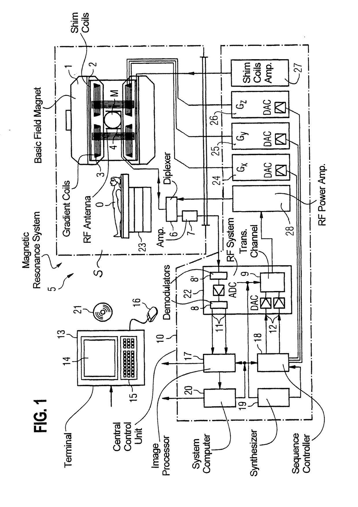
[0017]FIG. 1 schematically illustrates a magnetic resonance apparatus 5 (a magnetic resonance imaging or tomography device). A basic field magnet 1 generates, a temporally constant strong magnetic field for the polarization or alignment of the nuclear spin in a region of an examination subject O, such as a portion of a human body that is to be examined, lying on a table 23 in order to be moved into the magnetic resonance apparatus 5. The high degree of homogeneity in the basic magnetic field necessary for the magnetic resonance measurement (data acquisition) is defined in a typically sphere-shaped measurement volume M, in which the portion of the human body that is to be examined is placed. In order to support the homogeneity requirements temporally constant effects are eliminated by shim-plates made of ferromagnetic materials are placed at appropriate positions. Temporally variable effects are eliminated by shim-coils 2 and an appropriate control unit 27 for the shim-coils 2.

[0018]...


PUM


Abstract

In a magnetic resonance (MR) navigator-based method and apparatus, MR data are acquired from a large joint of a patient, which is not modelable as a whole based on a single rigid body model. The field of view which the MR data are acquired is divided in a processor into multiple sub-sections, with each sub-section being modelable based on a rigid body model. MR navigator signals are acquired from each of the sub-sections, and these navigator signals are used in a motion tracking algorithm that is based on a rigid body model in order to generate a modeling result that tracks the movement of the overall joint within the field of view. The modeling result can be used for prospective or retrospective motion correction of the MR data.

Description

BACKGROUND OF THE INVENTION[0001]Field of the Invention[0002]The present invention concerns methods and systems for magnetic resonance imaging, and in particular concerns methods and systems for implementing motion correction in magnetic resonance imaging of large joints, such as the knee.[0003]Description of the Prior Art[0004]Magnetic resonance (MR) imaging is a known technology with which images from the interior of an examination subject, such as a patient, can be generated. Simply described, the examination subject is placed in a magnetic resonance imaging scanner in which a strong, static, homogenous basic magnetic field is generated, typically having a field strength of 0.2 through 7 or more Tesla, causes nuclear spins in the subject to be oriented along the field lines of the basic magnetic field. In order to trigger magnetic resonance signals, the examination subject is irradiated with radio-frequency excitation pulses (RF pulses), that cause the nuclear spins to deviate fr...

Claims


Application Information

Patent Timeline
Patent Type & Authority Applications(United States)
IPC IPC(8): G01R33/483A61B5/055G01R33/565
CPCG01R33/4835A61B5/055G01R33/56509A61B5/0037A61B5/7207A61B5/748
Inventor BECK, THOMASBHAT, HIMANSHU
Owner SIEMENS HEALTHCARE GMBH
Why Eureka
Try Eureka