Method for measuring size of lesion which is shown by endoscope, and computer readable recording medium

a technology of endoscopy and measurement method, which is applied in the field of measuring the can solve the problems of not easy to grasp the exact size of the lesion and the exact opinion, and achieve the effect of accurate and rapid calculation, which is conducive to medical doctors in diagnosis and treatmen

Inactive Publication Date: 2017-06-01
PARK HYUN JUN +2
View PDF4 Cites 12 Cited by
  • Summary
  • Abstract
  • Description
  • Claims
  • Application Information

AI Technical Summary

Benefits of technology

[0019]The present invention has an advantageous effect in that size of the lesion can be accurately and quickly calculated from an image photographed by using a photographing device such as an endoscope, which is conducive to medical doctors in diagnosis and treatment.

Problems solved by technology

However, there are currently few methods for measuring an actual size of an image observed on an endoscope used in the clinic, and therefore, the current situation is that it is not easy to grasp the exact size of lesion and exact opinion.

Method used


Image

  • Method for measuring size of lesion which is shown by endoscope, and computer readable recording medium
  • Method for measuring size of lesion which is shown by endoscope, and computer readable recording medium
  • Method for measuring size of lesion which is shown by endoscope, and computer readable recording medium

Examples


Embodiment Construction

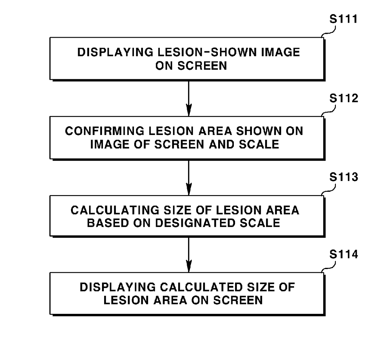
[0028]Hereinafter, example embodiments of the present invention will be described in more detail with reference to the accompanying drawings.

[0029]The method for measuring size of lesion which is shown by endoscope according to the present invention is performed by an image processing program performed by a computer system (device). At this time, the computer system may be any types of computers including personal computers, mobile computers, tablet computers, and diagnosis-dedicated terminals. Furthermore, the image processing program is defined by a computer program configured to execute the method for measuring size of lesion which is shown by endoscope according to the present invention regardless of its name. The image processing program may be variably embodied by being configured to be independently executed, by being configured as a module of another program, or being configured to be called by another program in a distributed format.

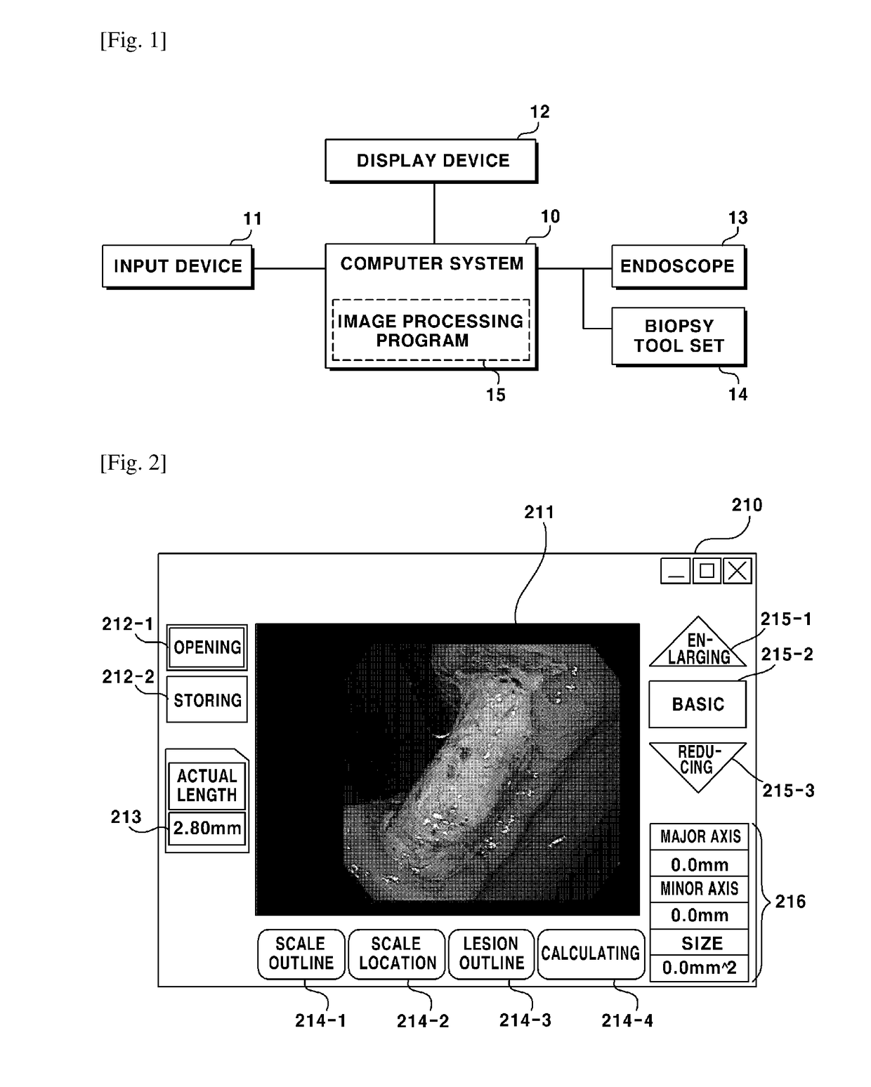
[0030]FIG. 1 is a schematic block diagram...


PUM


Abstract

A method is disclosed for measuring size of lesion from an image photographed by using a photographing device such as an endoscope. An image shown with lesion is shown on a screen, a lesion area is confirmed from the image shown on the screen, and the size of lesion area is calculated and displayed based on designated scale, whereby the size of lesion can be accurately provided, which is conducive to medical doctors in diagnosis and treatment.

Description

TECHNICAL FIELD[0001]The teachings in accordance with exemplary and non-limiting embodiments of this invention relate generally to a method for measuring size of lesion which is shown by endoscope, and more particularly, to a method for measuring size of lesion which is shown by endoscope, whereby the size of lesion can be accurately calculated and displayed from an image photographed by using a photographing device including an endoscope.BACKGROUND ART[0002]Absence and / or presence of diseases, and states of the diseases are ascertained using photographing devices including endoscopes in various medical fields. In general, information on size of lesion on an endoscope acts as an important factor in determining diagnosis and treatment direction of diseases. For example, cancer or adenoma found by an endoscope can be determined in treatment and method using the endoscope depending on sizes of the cancer and adenoma.[0003]When the exact sizes of various lesions after operations are kno...

Claims


Application Information

Patent Timeline
Patent Type & Authority Applications(United States)
IPC IPC(8): A61B5/107A61B1/04G06T7/60A61B5/00A61B10/04G06T7/00A61B1/00A61B5/103
CPCA61B5/1079A61B1/00009A61B1/0005A61B1/04A61B5/1032G06T2207/30096A61B5/743A61B10/04G06T7/0012G06T7/60G06T2207/10068A61B5/1076A61B5/748G06T7/62A61B5/103A61B5/107
Inventor PARK, HYUN JUNAHN, JI YONGLEE, JI SUK
Owner PARK HYUN JUN
Why Eureka
Try Eureka