Device, system and method for segmenting an image of a subject

a technology of image segmentation and subject, applied in the field of subject image segmentation devices, systems and methods, can solve the problems of difficult to achieve the technique, difficult to segment images, and difficult to achieve the effect of achieving the effect of improving the reliability of interactive image segmentation, increasing the efficiency of image segmentation, and increasing the accuracy of image segmentation

Active Publication Date: 2017-06-22
KONINKLJIJKE PHILIPS NV
View PDF2 Cites 7 Cited by
  • Summary
  • Abstract
  • Description
  • Claims
  • Application Information

AI Technical Summary

Benefits of technology

[0022]In another preferable embodiment, the gradient peak comprises a maximum gradient peak and / or a first gradient peak beginning from said image control point. The maximum gradient peak is understood as obtained by finding the maximum value of the image gradient detected all over the volume and / or the path. In this way, the target point can be located at the boundary between two different material and / or tissue types with high accuracy. This advantageously leads to increased reliability of the interactive image segmentation. Further, the present invention takes into account that the distance between the first contour and the structure of the subject is relatively small in certain regions of the first contour, in particular in the HARs. For such regions, it is very likely that only one gradient peak is present within a short distance from the corresponding image control point. Therefore, it is sufficient to detect the first gradient peak in order to detect a maximum gradient peak or a gradient peak close to the maximum gradient peak in its magnitude and / or position. Advantageously, the efficiency of the image segmentation is further increased.
[0023]In another preferable embodiment, the segmentation unit is configured to identify the target points only if the gradient peak is above a predefined threshold gradient. It can be prevented in this way that target points that are not qualified for reasonably defining the second contour are identified. Advantageously, the reliability of image segmentation is further increased.
[0024]In another preferable embodiment, the second contour comprises a distance to the first contour increasing with the motion parameter. A band is formed in this way as being defined by the first and the second contour, wherein the bandwidth is larger in the LARs than in the HARs. Such a band advantageously serves as a refined starting point for the graph cut, instead of a randomly chosen band, thus leading to increased reliability of the segmentation result.
[0025]In another preferable embodiment, the image comprises a first and a second image slice, further comprising a transferring unit for transferring the structure and / or the first contour and / or the second contour from the first to the second image slice. In this way, the structure and / or and first contour and / or the second contour can be used to segment the second image slice. This advantageously improves the image segmentation by adjusting the first and / or the second contour or by predicting a new contour.
[0026]In another preferable embodiment, the image control point unit is configured to distribute a plurality of additional image control points without using the motion parameter. In particular, the path corresponding to the additional image control points comprises a length correlated with a curvature of the structure. Advantageously, the present invention enables both interactive image segmentation and automatic image segmentation and a combination of both, leading to increased user flexibility.
[0027]In another preferable embodiment, the motion parameter registering unit is configured to signal an error when said motion parameter is below a predefined threshold parameter. The present invention enables to indicate a segmentation result that is counter-intuitive to the image content, for instance due to the presence of implants, or to indicate the case where no image gradient can be measured. Advantageously, this leads to a more secure and reliable image segmentation.

Problems solved by technology

However, this technique is sometimes unattainable due to limitations such as image acquisition errors, abnormalities in the image content and the presence of local image ambiguities.
However, the approaches known in the art are limited in their accuracy and efficiency since the information about user-initiated motions is not effectively applied to speed up a process of image segmentation.
In addition, the delineation of structures on 3D medical images slice by slice is tedious and time consuming.
Some smart tools snap to image gradients, but the delineation still requires a lot of precise mouse movements.
Often, this is done manually and the user has to outline the structure carefully with a contouring tool, which is very time consuming.
However, this strategy only works for very few structures.
Further, many algorithms require prior knowledge on structures to contour from a library.
Thus, for many known structures and all new or uncommon structures, still a lot of time is needed for accurate delineation.

Method used


Image

  • Device, system and method for segmenting an image of a subject
  • Device, system and method for segmenting an image of a subject
  • Device, system and method for segmenting an image of a subject

Examples


Embodiment Construction

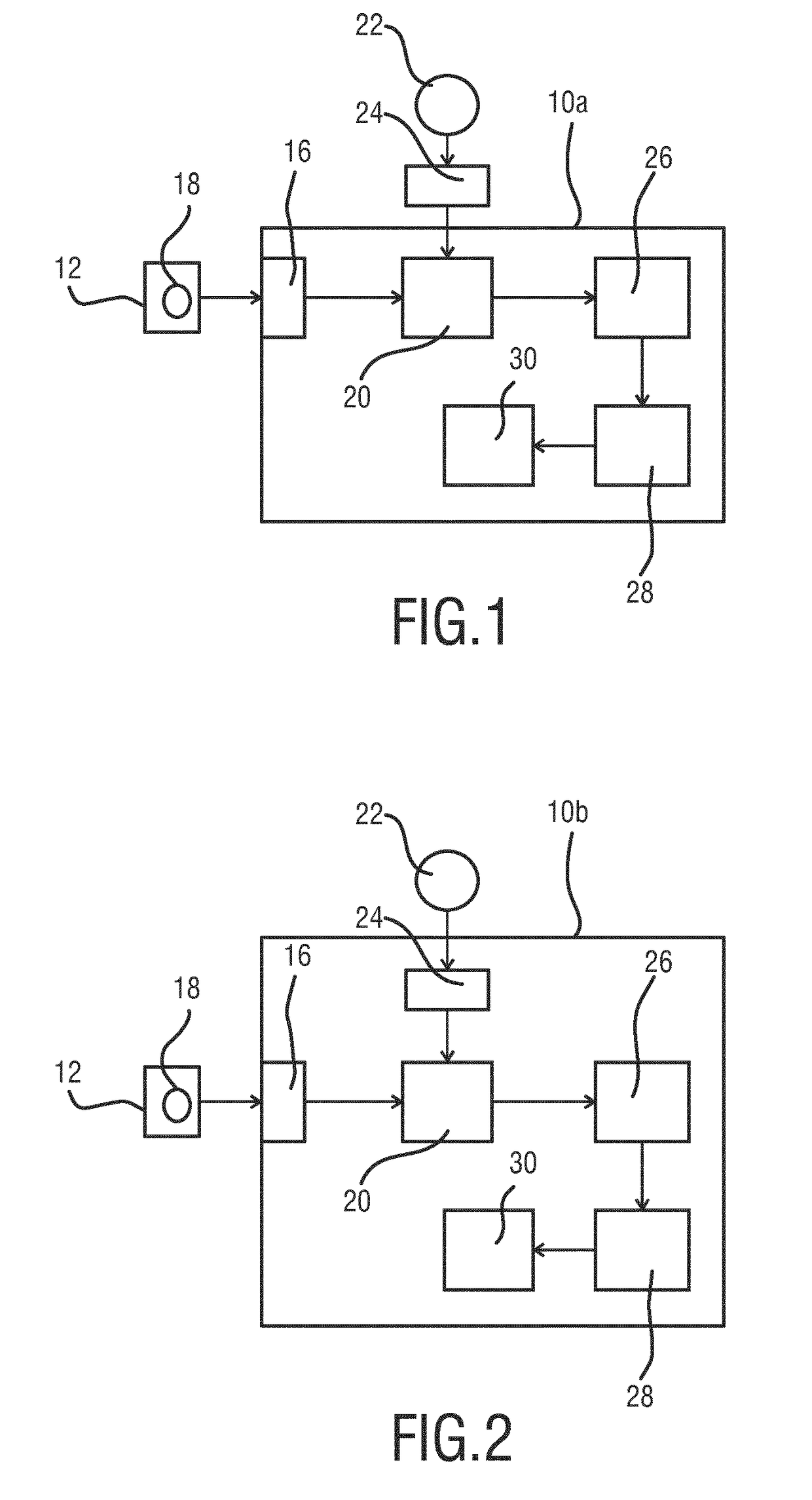
[0035]With reference to FIG. 1, a schematic block diagram of the device 10a for segmenting an image 12 of a subject in accordance with a first embodiment is shown. The device 10a comprises a data interface 16 for receiving the image 12 of the subject. The image 12 comprises a structure 18 which corresponds to a part of the subject. The subject is typically a living being such as a human, wherein the part to which the structure 18 corresponds may e.g. comprise an anatomical structure such as the lung, the brain, the heart, the stomach, etc. It is understood by the person skilled in the art that the structure 18 is enclosed by a boundary separating the structure 18 from the image content of the image 12 outside the structure 18.

[0036]The data interface 16 may be any type of data interface known in the art, in particular a data connection between an imaging apparatus to the device 10a. Such a data connection serves to transfer image data from the imaging apparatus to the device 10a for...


PUM


Abstract

The present invention relates to a device for segmenting an image of a subject (36), comprising a data interface for receiving an image of said subject (36), said image depicting a structure of said subject (36), a translation unit for translating a user-initiated motion of an image positioner means into a first contour (38) surrounding said structure, a motion parameter registering unit for registering a motion parameter of said user-initiated motion to said first contour (38), said motion parameter comprising a speed and/or an acceleration of said image positioner means, an image control point unit for distributing a plurality of image control points (40) on said first contour with a density decreasing with said motion parameter, and a segmentation unit for segmenting said image by determining a second contour (44) within said first contour based on said plurality of image control points (40), said segmentation unit being configured to use one or more segmentation functions.

Description

FIELD OF THE INVENTION[0001]The present invention relates to a device, system and method for segmenting an image of a subject. It finds application in medical imaging, in particular for diagnosis or treatment planning.BACKGROUND OF THE INVENTION[0002]In medical imaging, image segmentation is one of the most important tasks of surgical persons to obtain in-depth analysis of medical images. The goal of image segmentation is to identify a region of interest (ROI) and to highlight the boundary of the ROIs so that an operator carrying out image analysis is able to tell the ROIs from the rest of the image content.[0003]There exist numerous tours for image segmentation. For instance, automatic processing is advantageous when it is applied to well defined structures and standard contouring definitions. However, this technique is sometimes unattainable due to limitations such as image acquisition errors, abnormalities in the image content and the presence of local image ambiguities. In order...

Claims


Application Information

Patent Timeline
Patent Type & Authority Applications(United States)
IPC IPC(8): G06T7/12G06T7/149G06T7/00
CPCG06T7/12G06T7/0012G06T2207/30004G06T2207/20104G06T2207/20116G06T7/149G06T2207/20168G06T2207/30008
Inventor SCHADEWALDT, NICOLESCHULZ, HEINRICHBYSTROV, DANIELFRANZ, ASTRID RUTH
Owner KONINKLJIJKE PHILIPS NV
Why Eureka
Try Eureka