Deep brain source imaging with m/eeg and anatomical MRI

a brain source and anatomical technology, applied in the field of deep brain source imaging with m/eeg and anatomical mri, can solve the problems of high invasiveness, high invasiveness, and difficulty in technology using known methods, and achieve the effect of lower resolution and higher resolution

Active Publication Date: 2017-11-23
THE GENERAL HOSPITAL CORP +1
View PDF8 Cites 7 Cited by
  • Summary
  • Abstract
  • Description
  • Claims
  • Application Information

AI Technical Summary

Benefits of technology

[0012]The step of constructing hierarchical source spaces can comprise (a) grouping clusters of neocortical dipoles and hippocampal dipoles into surface patches, and (b) grouping clusters of subcortical dipoles into volume subdivisions, wherein the patches and the subdivisions each vary in levels of coarseness across the hierarchies. The step of constructing patches and subdivisions can include sizing the patches and subdivisions to homogenize current strengths, and can also include assigning higher resolution to regions with high current densities and lower resolution to regions with low current densities, thereby enabling weaker and stronger gain regions to have comparable consideration in an inverse solution.

Problems solved by technology

Understanding human brain function in cognition, behavior and illness remains an enduring challenge.
Thus, many scientific challenges in understanding brain function come about from the need to parse these complex dynamical interactions across diverse spatial and temporal scales, such as across superficial cortical and deeper subcortical regions, and on time scales ranging from milliseconds to seconds and minutes.
Monitoring these complex dynamic interactions at the relevant spatial and temporal scales, and as they occur in normal, clinical and disease states is, however, technologically challenging using known methods.
These techniques can be either invasive or non-invasive.
While these techniques provide high spatial and temporal resolution of regional neuronal dynamics, they are highly invasive, and thus are typically applied in critical clinical settings (such as during surgery).
This limits the types of patients / subjects, cognitive tasks, and behaviors that can be monitored with such techniques.
Further, invasive electrophysiological techniques are limited to the focal regions wherein recording electrodes are placed (typically superficial cortical areas).Therefore, these techniques have limited spatial span for characterizing deep subcortical regions that have critical roles in a variety of cognitive and clinically relevant brain states.
However, the data do not directly pinpoint spatial locations or regions where the neuronal currents originate.
However, because M / EEG signal amplitudes attenuate steeply with increasing source-sensor distance, it has been challenging to use electromagnetic source imaging for assessing neuronal dynamics in deeper subcortical regions that are farther away from non-invasive M / EEG sensors.
Overall, although existing electrophysiology-based techniques provide excellent temporal resolution, they have limited spatial resolution for activity in deep brain regions.

Method used


Image

  • Deep brain source imaging with m/eeg and anatomical MRI
  • Deep brain source imaging with m/eeg and anatomical MRI
  • Deep brain source imaging with m/eeg and anatomical MRI

Examples


Embodiment Construction

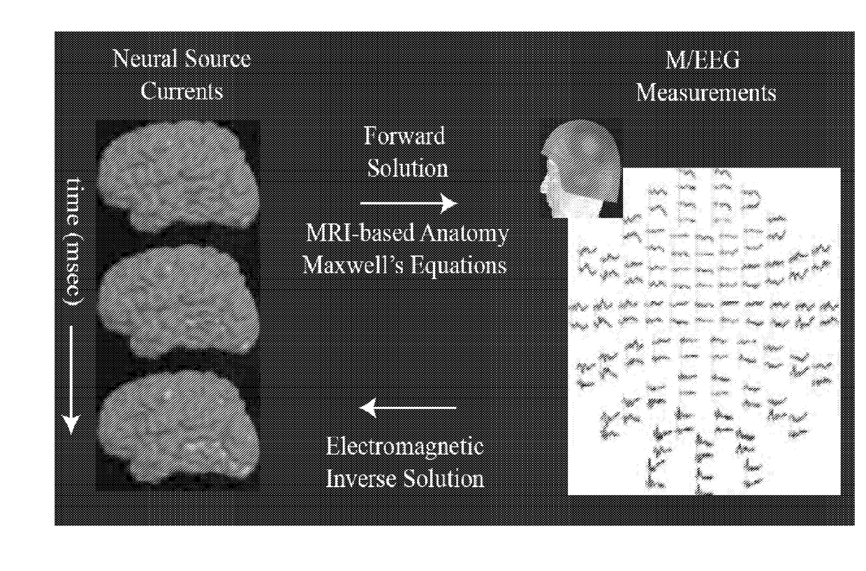
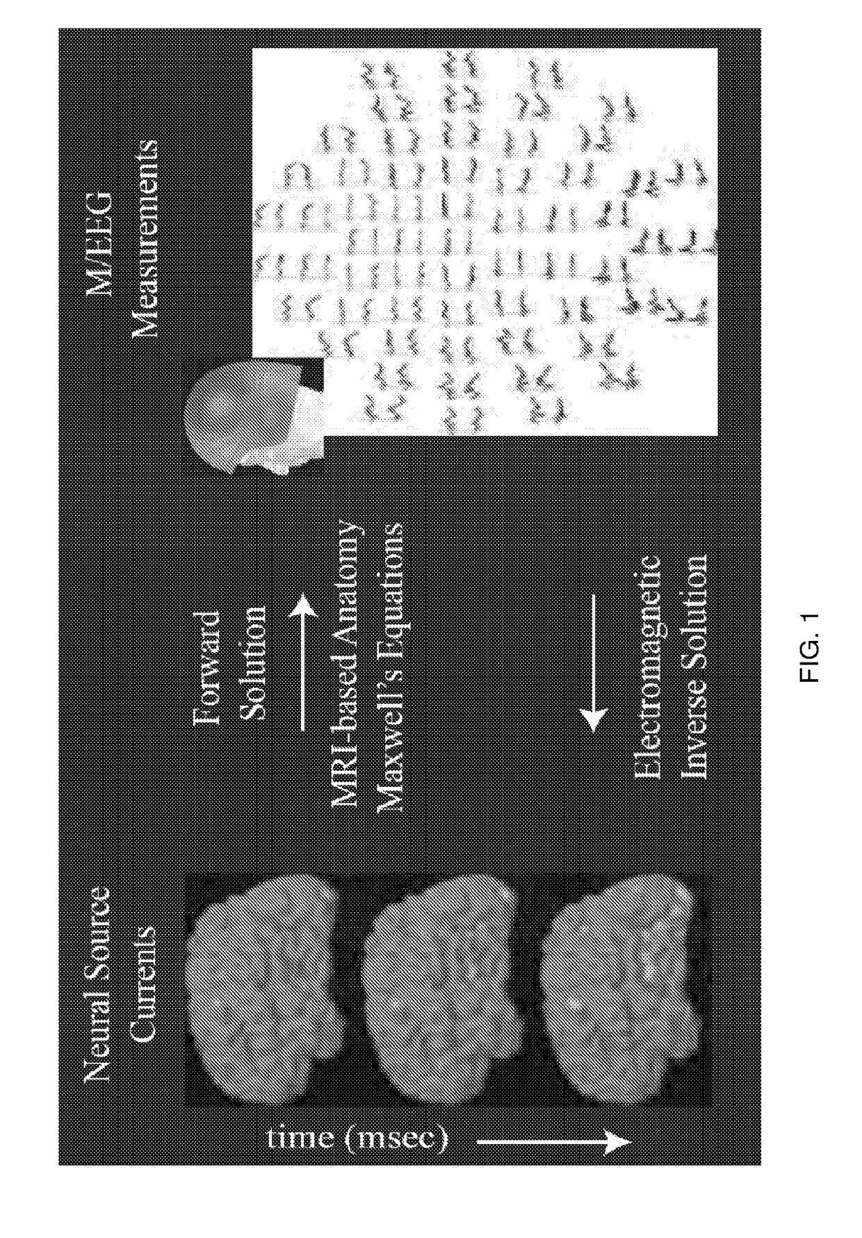
[0030]The present disclosure describes methods of electromagnetic source imaging with non-invasive M / EEG recordings and MRI-based anatomic measures that can be used to acquire gain-insensitive information in M / EEG arising from subcortical and cortical structures, and provides a hierarchical subspace pursuit algorithm to estimate neural currents in the subcortical structures. Distinctions between field patterns can be used to localize subcortical sources and distinguish subcortical versus cortical contributions using the hierarchical subspace pursuit process or algorithm.

[0031]Referring now to FIG. 2, a flowchart illustrates a series of steps for estimating source currents from deep brain regions with a hierarchical subspace pursuit algorithm. Initially, M / EEG data are acquired during a neurophysiologic paradigm of interest. These recordings are complemented with magnetic resonance images of the subject's brain anatomy (step 10). Next, distributed source spaces are constructed from t...


PUM


Abstract

A method for non-invasively resolving electrophysiological activity in sub-cortical structures located deep in the brain by comparing amplitude-insensitive M/EEG field patterns arising from activity in subcortical and cortical sources under physiologically relevant sparse constraints is disclosed. The method includes a sparse inverse solution for M/EEG subcortical source modeling. Specifically, the method employs a subspace-pursuit algorithm rooted in compressive sampling theory, performs a hierarchical search for sparse subcortical and cortical sources underlying the measurement, and estimates millisecond-scale currents in these sources to explain the data. The method can be used to recover thalamic and brainstem contributions to non-invasive M/EEG data, and to enable non-invasive study of fast timescale dynamical and network phenomena involving widespread regions across the human brain.

Description

CROSS-REFERENCE TO RELATED APPLICATIONS[0001]This application claims the benefit of U.S. Provisional patent application Ser. No. 62 / 076,525 filed on Nov. 7, 2014, which is incorporated herein by reference.BACKGROUND[0002]Understanding human brain function in cognition, behavior and illness remains an enduring challenge. This is because the brain is highly complex, with billions of neurons, interacting dynamically to receive, process, retrieve, transmit, and store information. Neurons signal via millisecond electrical impulses, communicate with other neurons in both local (mm-range) and distant (cm-range) regions. These interactions evolve instantaneously in response to external stimuli, drugs, or feedback from other brain regions, during creation and expression of memory, emotion and perception, across states of arousal, and over time due to plasticity, learning, development and aging. Thus, many scientific challenges in understanding brain function come about from the need to parse...

Claims


Application Information

Patent Timeline
Patent Type & Authority Applications(United States)
IPC IPC(8): A61B5/0436A61B5/0476A61B5/055A61B5/00A61B5/336
CPCA61B5/0436A61B5/4064A61B5/055A61B5/0476A61B5/336A61B5/369
Inventor KRISHNASWAMY, PAVITRAPURDON, PATRICKOBREGON-HENAO, GABRIELHAMALAINEN, MATTIBABADI, BEHTASH
Owner THE GENERAL HOSPITAL CORP
Why Eureka
Try Eureka