Automated delineation of nuclei for three dimensional (3-d) high content screening

a three-dimensional, high-content technology, applied in the field of system and method for image processing, can solve the problems of slow and unreliable 3-d image processing, negatively affecting the performance of studying a drug response,

Active Publication Date: 2018-03-15
CEDARS SINAI MEDICAL CENT
View PDF5 Cites 12 Cited by
  • Summary
  • Abstract
  • Description
  • Claims
  • Application Information

AI Technical Summary

Benefits of technology

[0004]In various embodiments, the invention teaches a method that includes delineating a nucleus of one or more cells in an image of one or more cells by utilizing a mathematical algorithm that is sensitive to the spherical shape of a nucleus of one or more cells in the image. In some embodiments, the method further includes positioning a seed within one or more cell nucleus in the image. In certain embodiments, the seed is obtained through a radial symmetry image that is a weighted sum of orientation projection and magnitude projection images derived from local image gradients. In some embodiments, the method further includes applying watershed-based segmentation in which the positions of two or more seeds is considered during segmentation. In certain embodiments, one or more of the one or more cells have been stained with one or more markers. In some embodiments, one or more of the markers are fluorescent biomarkers. In certain embodiments, one or more of the fluorescent biomarkers are immunofluorescent biomarkers. In some embodiments, at least one cell in the image has been immunolabeled with a 5-methylcytosine antibody and/or 4′6-diamidino-2-phenylindole (DAPI). In certain embodiments, the image is acquired by confocal microscopy. In some embodiments, the image includes cells from a preparation of a tissue mounted on a slide. In some embodiments, at least one cell in the image is a cancer cell or a pre-cancerous cell. In certain embodiments, at least one cell in the image has been treated with a drug. In some embodiments, the drug is a chemotherapeutic dr

Problems solved by technology

However, cell confluency, complexity of nuclear staining as well as poor contrast between nuclei and background result

Method used


Image

  • Automated delineation of nuclei for three dimensional (3-d) high content screening
  • Automated delineation of nuclei for three dimensional (3-d) high content screening
  • Automated delineation of nuclei for three dimensional (3-d) high content screening

Examples


example 1

[0043]By way of additional background, the objective and quantitative analysis of images from cells labeled with immunofluorescent probes takes microscopy from being purely visual, qualitative and subjective to a higher level. Quantitative analysis of fluorescently labeled molecular targets in cells can provide information of their spatial distribution, amounts and topology. The advantage of quantitative microscopy in cellular studies is further enhanced by the addition of automation. High-content screening (HCS)—or the automated acquisition of fluorescently labeled cellular images followed by an automated analysis enables the quantitative assessment of large numbers of images, thereby paving the way for large scale experiments in which multiple conditions are examined simultaneously, as described in Cox K L et al. Immunoassay Methods. In: Assay Guidance Manual. Edited by Sittampalam G S, et al. Bethesda (Md.); 2004; Buchser W, et al.: Assay Development Guidelines for Image-Based Hi...

example 2

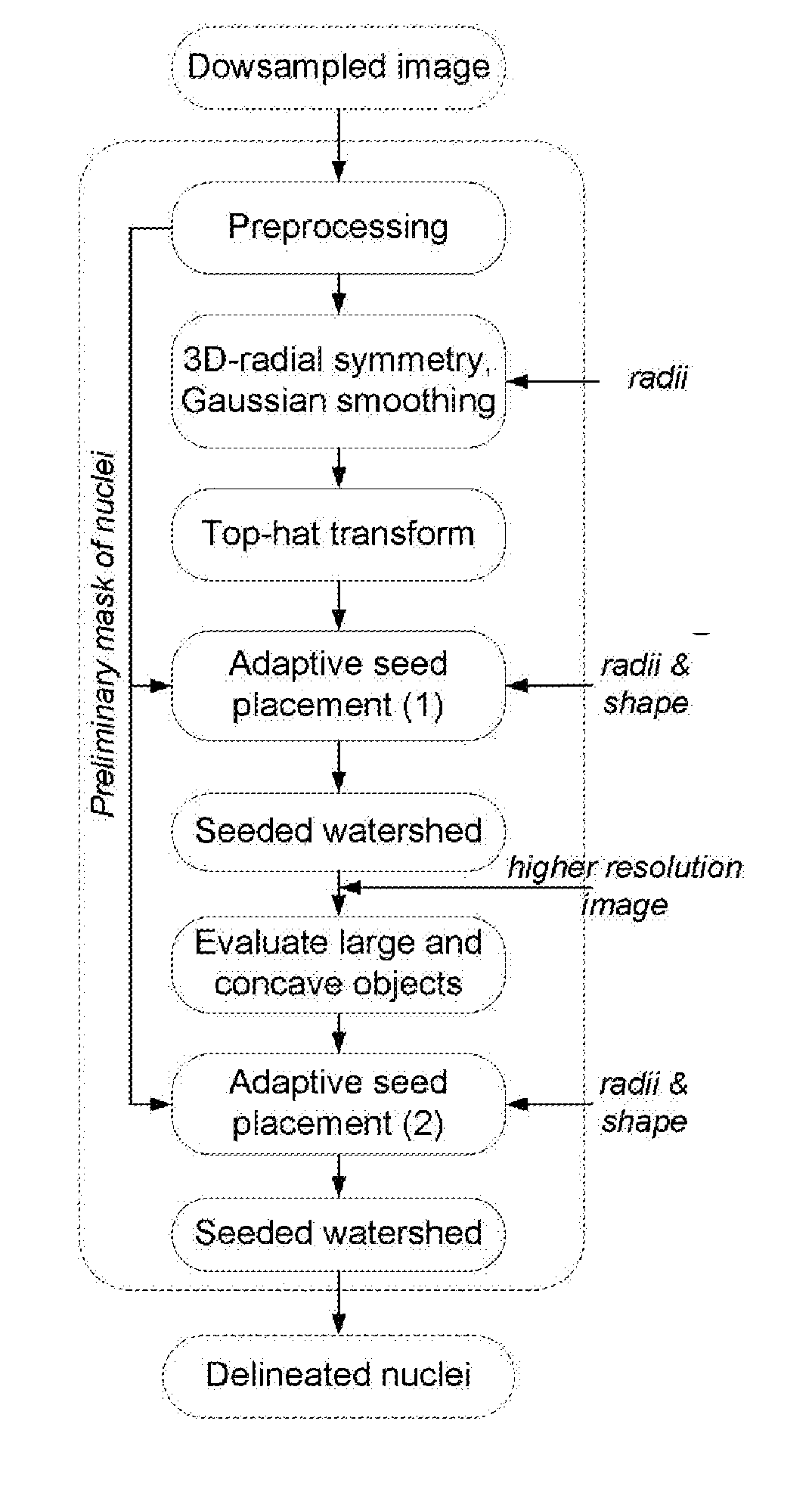
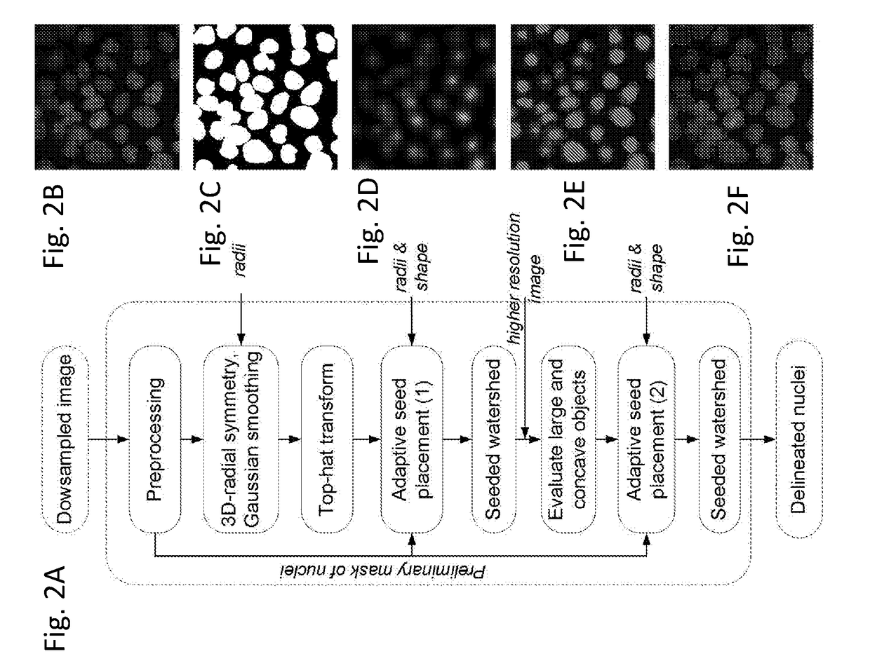
[0076]The results from another approach to delineating cell nuclei are demonstrated in FIGS. 11 and 12. In this approach two-dimensional color images of human tissues stained with hematoxylin and eosin were color-normalized and then color-deconvoluted to provide a monochromatic hematoxylin image for the segmentation of nuclei. Similarly to the 3D-RSD, a set of parameters namely the nuclear radii Riε[5, 11] and nuclear shape defined by shullxy=0.95 and exy=1.5 that was a priori defined. The nuclear mask was obtained by a parameter free histogram thresholding (see Zack G W, et al.: Automatic measurement of sister chromatid exchange frequency. The journal of histochemistry and cytochemistry: official journal of the Histochemistry Society 1977, 25(7):741-753). The segmentation was then performed using the following sequence of routines: 2-D radial symmetry transform followed by the 2-D top-hat transform to obtain a radial symmetry image, and the adaptive seed placement followed by the 2...


PUM


Abstract

In various embodiments, the invention teaches systems and methods for analyzing an image of cells, including an image obtained by confocal microscopy, and delineating cell nuclei in the image. In some embodiments, the systems and methods apply a three-dimensional (3-D) radial symmetry transform followed by adaptive post processing of symmetry images to arrive at a mask of seeds that can guide watershed-based segmentation.

Description

GOVERNMENT RIGHTS[0001]This invention was made with government support under grant CA143618 awarded by the National Institutes of Health. The government has certain rights in the invention.FIELD OF THE INVENTION[0002]The present invention generally relates to systems and methods for image processing.BACKGROUND[0003]High-resolution three-dimensional (3-D) microscopy combined with multiplexing of fluorescent labels allows high-content analysis of large numbers of cell nuclei. The full automation of 3-D screening platforms necessitates image processing algorithms that can accurately and robustly delineate nuclei in images with little to no human intervention. Imaging-based high-content screening was originally developed as a powerful tool for drug discovery. However, cell confluency, complexity of nuclear staining as well as poor contrast between nuclei and background result in slow and unreliable 3-D image processing and therefore negatively affect the performance of studying a drug r...

Claims


Application Information

Patent Timeline
IPC IPC(8): G06K9/00G01N1/30G01N33/50
CPCG06K9/0014G01N1/30G01N33/5011G06V20/695
Inventor GERTYCH, ARKADIUSZKNUDSEN, BEATRICE
Owner CEDARS SINAI MEDICAL CENT
Why Eureka
Try Eureka