In vitro artificial lymph node method for sensitization and expansion of t cells for therapy and epitope mapping

a technology of t cell sensitization and epitope mapping, applied in the direction of antibody medical ingredients, instruments, drug compositions, etc., can solve the problems of loss of antigen specificity and/or function, poor expansion level, and premature activation-induced cell death (apoptosis)

Inactive Publication Date: 2018-06-21
THE TRUSTEES OF THE UNIV OF PENNSYLVANIA
View PDF0 Cites 0 Cited by
  • Summary
  • Abstract
  • Description
  • Claims
  • Application Information

AI Technical Summary

Benefits of technology

[0018]c) subsequently contacting the stimulated antigen specific T cell with IL-2, thereby generating an expanded antigen specific T cell population that maintains antigen specificity and cellular function.
[0029]c) subsequently contacting the stimulated antigen specific T cell with IL-2, thereby generating an expanded antigen specific T cell population that maintains antigen specificity and cellular function.

Problems solved by technology

However, in practice, many technical problems exist including poor levels of expansion, premature activation-induced cell death (apoptosis), or loss of antigen specificity and / or function.
Part of the problem lies in the inability to replicate, in vitro, the environment inside the body where antigen-specific T cell expansion occurs, which is the lymph node.

Method used


Image

  • In vitro artificial lymph node method for sensitization and expansion of t cells for therapy and epitope mapping
  • In vitro artificial lymph node method for sensitization and expansion of t cells for therapy and epitope mapping
  • In vitro artificial lymph node method for sensitization and expansion of t cells for therapy and epitope mapping

Examples


example 2

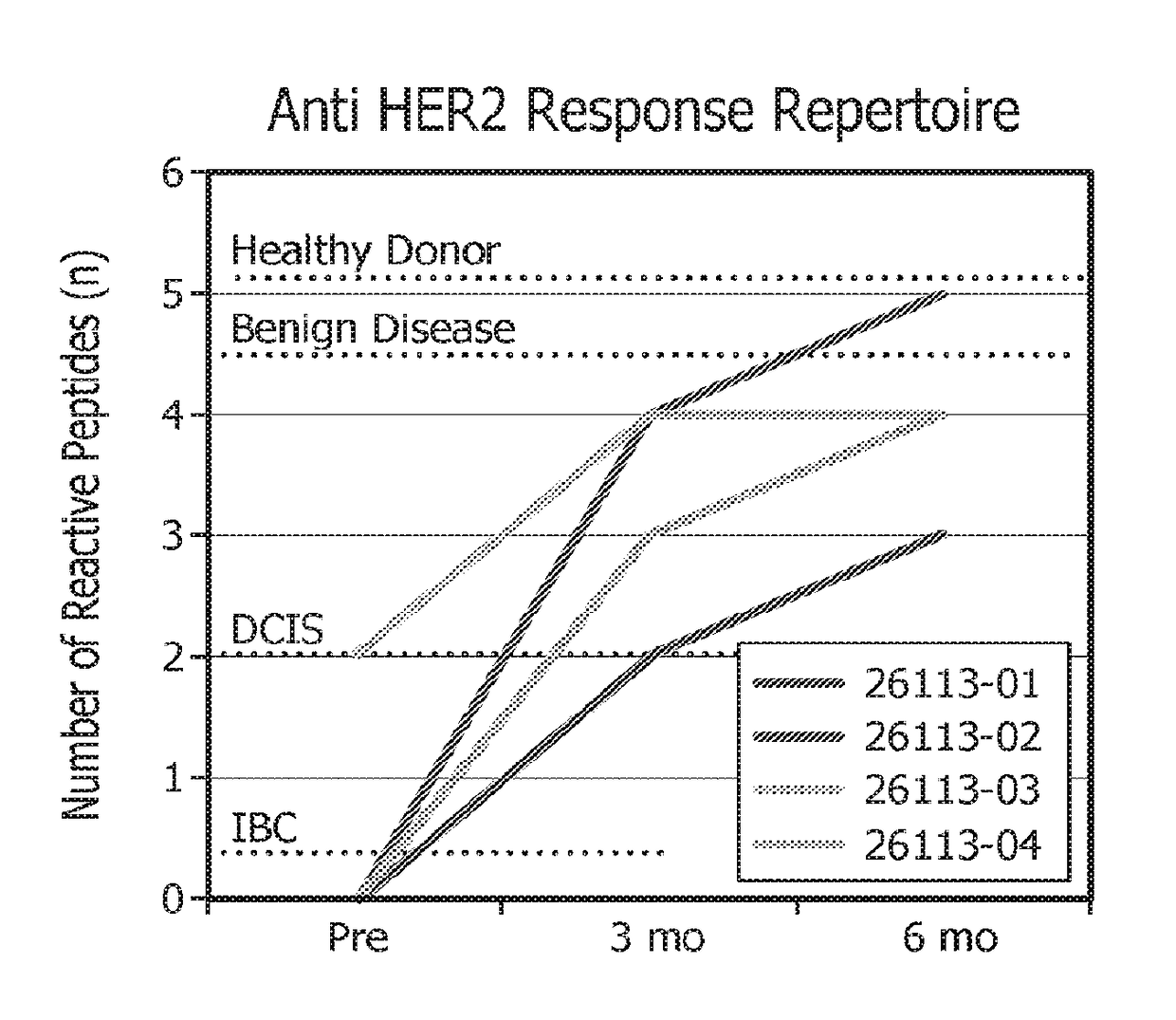
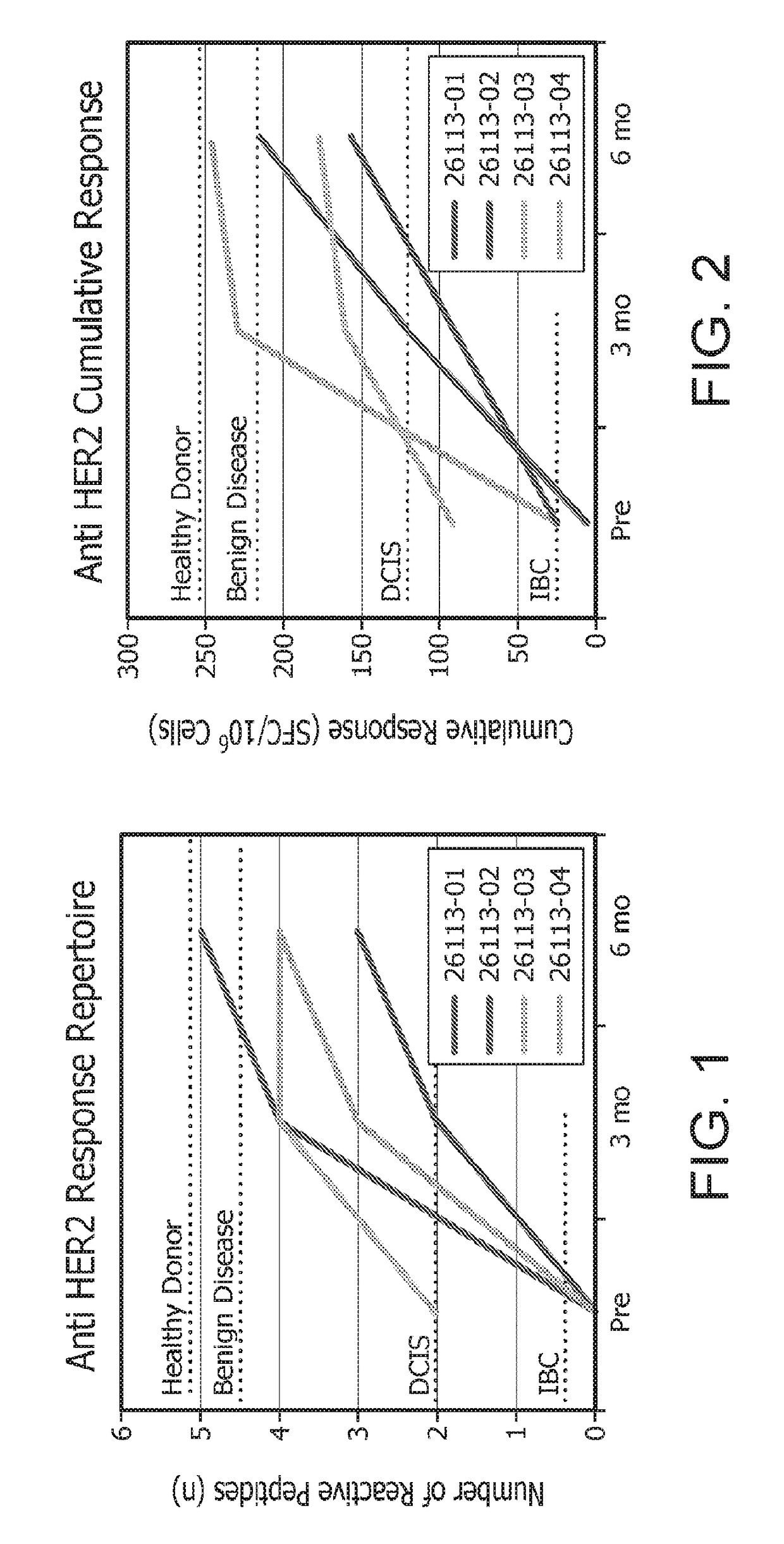
Expansion of HER2-Specific Th1 Cells

[0219]The following studies were designed to explore the role of adoptive T-cell transfer in restoring anti-HER2 Th1 immunity. T cells are expanded to a level necessary for adoptive therapy and epitope mapping studies while maintaining antigen specificity and cellular function.

[0220]Briefly, in vitro, HER2-specific Th1 cells were generated by co-culture with HER2-peptide pulsed DC1s and expanded using IL-2 alone or IL-2, IL-7, and IL-15. Th1 cells were subsequently expanded either by repeat HER2-peptide pulsed DC1 co-culture or via anti-CD3 / CD28 stimulation. Fold expansion was defined as: (#T-cells post expansion / #T-cells pre expansion); specificity was measured by antigen specific IFNγ production by ELISA.

[0221]As will be shown herein, repeated co-culture of CD4+ T cells with HER2 peptide pulsed DC1s stimulated with IL-2, IL-7, and IL-15 results in a significant expansion of highly specific anti-HER2 Th1 cells, providing a potential population of...

example 3 -

Example 3-Method for Expanding T Cells

[0223]HER2 Specific DC1 Preparation:

[0224]DC precursors were obtained from HER2 breast cancer patients (DCIS) who were vaccinated with HER2 peptide-pulsed DC1 vaccines, as described previously. DC precursors were obtained via tandem leukapheresis / countercurrent centrifugal elutriation. DCs were incubated at 3×10⁶ cells in 1 ml Macrophage Serum-free Medium (SFM-Gibco Life Technologies, Carlsbad, Calif.) with GM-CSF 50 ng / ml (Berlex, Richmond, Calif.) at 37° C. DCs were pulsed with a single HER2 peptide antigen (42-56, 98-114, 328-345, 776-790, 927-941, 1166-1180 (SEQ ID NOS 1-6)); 20 μg / ml) 48-72 hrs after the cells were initially plated. For maturation, DCs were further activated 6 hours later with IFN-γ (1000 U / ml) and the following day with lipopolysaccharide (“LPS”) (10 ng / ml). HER2 specific DC1s were harvested 6 hours after LPS administration at the point of maximum IL-12 production.

[0225]CD4+ T Cell Preparation:

[0226]Lymphocytes were also o...


PUM

PropertyMeasurementUnit
culture timeaaaaaaaaaa
temperatureaaaaaaaaaa
volumeaaaaaaaaaa

Abstract

A method of creating a microenvironment for culture expansion of T cells. The expanded T cells can be used for a variety of therapeutic and research purposes.

Description

[0001]This application claims priority and benefit from U.S. Provisional Patent Application Ser. No. 62 / 138,969 filed on Mar. 26, 2015.FIELD[0002]The present embodiments are directed to, in vitro artificial lymph node method for sensitization and expansion of T cells for therapy and epitope mapping and diagnostic monitoring methods, treatment methods and tools based thereon.BACKGROUND[0003]The lifetime risk of breast cancer development is nearly one in eight. The erb-B2 oncogene (HER-2 / neu) is a molecular driver that is overexpressed in a significant number of breast, ovarian, gastric esophageal, lung, pancreatic, prostate and other solid tumors. HER2 overexpression (“HER2pos”), a molecular oncodriver in several tumor types including approximately 20-25% of breast cancers (Meric, F., et al., J. Am Coll. Surg. 194:488-501 (2002)), is associated with an aggressive clinical course, resistance to chemotherapy, and a poor overall prognosis in breast cancer (“BC”). See, Henson, E. S., Cli...

Claims


Application Information

Patent Timeline
Patent Type & Authority Applications(United States)
IPC IPC(8): C12N5/0783G01N33/574A61K39/00
CPCC12N5/0636G01N33/574A61K39/0011A61K2039/5158A61K2039/55533A61K2039/5154C12N2501/11C12N2502/1121A61P35/00
Inventor CZERNIECKI, BRIAN J.LOWENFELD, LEA
Owner THE TRUSTEES OF THE UNIV OF PENNSYLVANIA
Why Eureka
Try Eureka