
Epitope mapping method

a mapping method and epitope technology, applied in the field of epitope characterization and vaccine design, can solve the problems of requiring a considerable amount of pre-existing knowledge to interpret sequencing data, limiting the rate of the iterative vaccine development approach, and considerable limitations in the method

Active Publication Date: 2022-01-04
THE SCRIPPS RES INST
View PDF0 Cites 0 Cited by
  • Summary
  • Abstract
  • Description
  • Claims
  • Application Information

AI Technical Summary

Benefits of technology

The patent describes a method for mapping the immune response to an immunogen by measuring differences in structural images of immune complexes formed between the immunogen and serum antibodies at different time intervals. The method allows for the simultaneous visualization of multiple antibodies targeting different epitopes in the immunogen. The immunogen can be a recombinant immunogen or an epitope. The method can be used to characterize the epitopes recognized by the antibodies and to improve the immunogen. The patent also describes a method for forming immune complexes and quantifying the specific immunoglobulin content in the complexes. Overall, the method provides a better understanding of the immune response to immunogens and can be used in the development of vaccines and therapeutic antibodies.

Problems solved by technology

One of the rate-limiting steps in the iterative vaccine development approach is in analysis of the polyclonal immune response elicited by immunization.
Recently methods such as use of deep sequencing of the B cell receptor (BCR) repertoire of responding B cells have been introduced; however, this method has considerable limitations because the heavy-light chain pairing is usually lost.
Similarly, next generation sequencing (NGS) analyses can be used to study responses but it requires a considerable amount of pre-existing knowledge to interpret the sequencing data.
This process is time consuming and limited to a relatively small number of samples.
Such analyses typically focus on antibodies with a biological function (e.g. neutralization) and often leave the remainder of the humoral immune response less well investigated.
Due to the high cost and labor involved, unsuccessful outcomes of vaccination experiments are hardly ever analyzed in detail, and the reasons for failure of a given immunogen to generate neutralizing response continues to remain unknown.

Method used


Image

  • Epitope mapping method
  • Epitope mapping method
  • Epitope mapping method

Examples


example 1

Generally

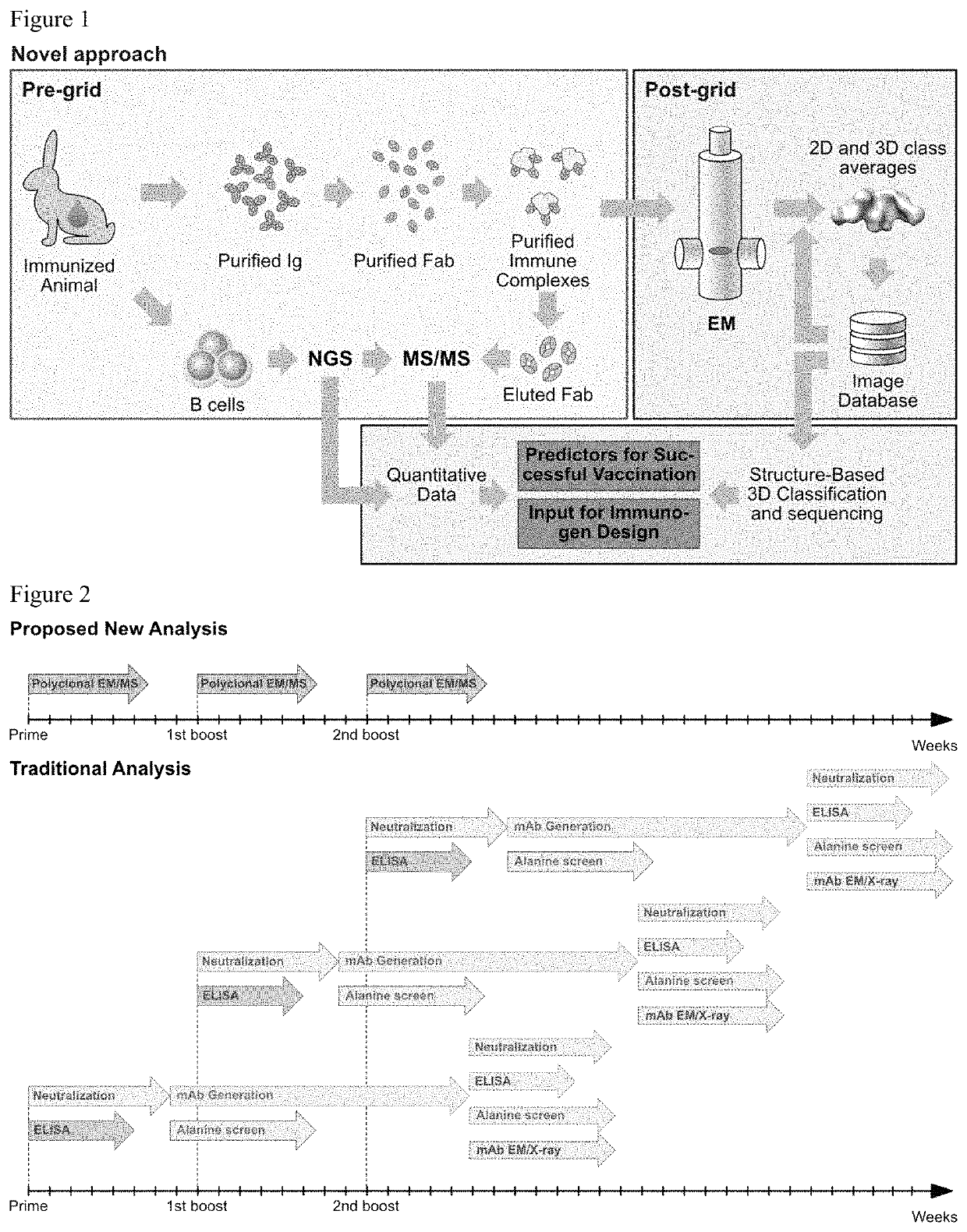
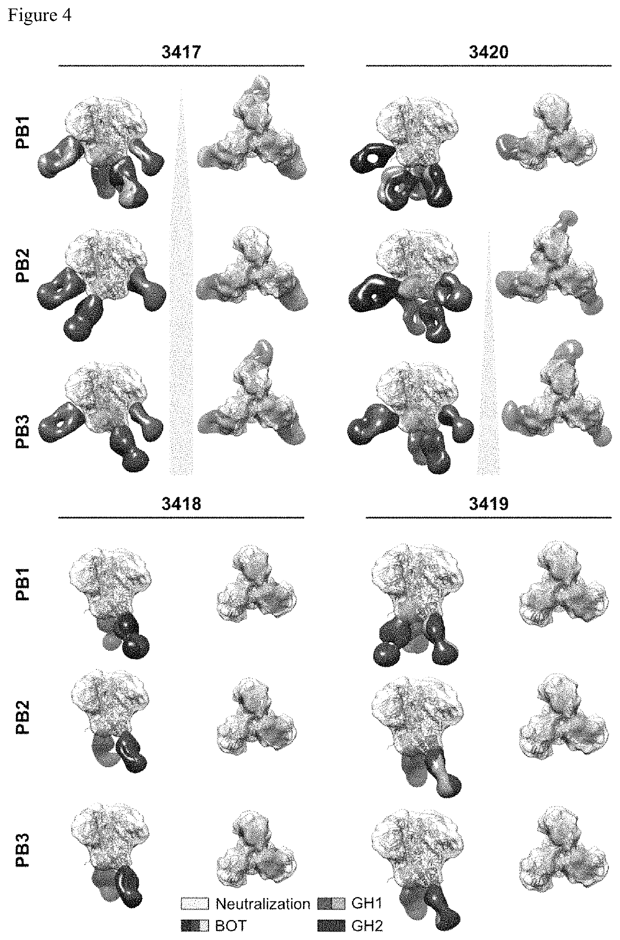
[0036]Polyclonal humoral immune responses, for example to natural infection and vaccination, are inherently complex and difficult to characterize using currently available methodologies. Mapping of all epitopes in an immune response is typically incomplete and, therefore, creates a barrier to fully understand the humoral response to infection and hence can hinder rational vaccine design efforts. The present disclosure provides a new methodology that the inventors have applied to characterize polyclonal responses in rabbits to immunization with a trimeric HIV-1 envelope glycoprotein subunit vaccine candidate, BG505 SOSIP.664, using single particle electron microscopy. This approach was able to detect known epitopes and reveal how antibody responses evolved during the prime-boost-boost-boost strategy that ultimately resulted in a neutralizing antibody response. Furthermore, previously unknown epitopes were uncovered, including non-neutralizing epitopes, as well as an epitope ...

example 2

BG505 SOSIP.664 Immunization

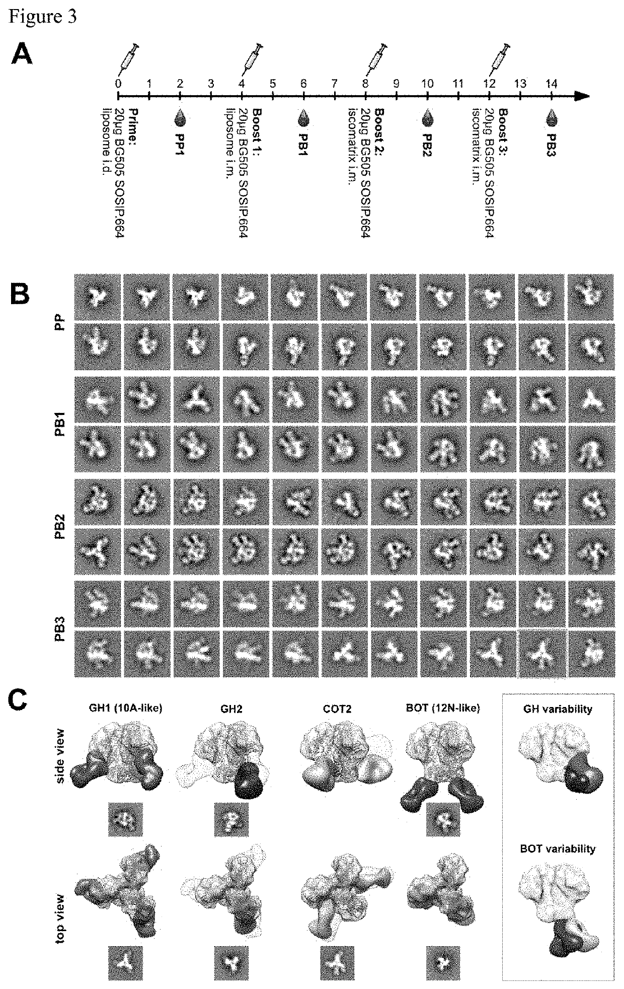
[0040]In one embodiment, sera from four BG505 SOSIP.664 immunized rabbits were used, that have been extensively characterized and disclosed by the authors in McCoy et al, Cell Rep. 2016 Aug. 30; 16(9): 2327-2338, and that are referred to as 3417, 3418, 3419, and 3420. Rabbits were immunized three times with BG505 SOSIP.664 and bled two weeks following each immunization (FIG. 3A). Sera obtained are referred to as PP, PB1, PB2, and PB3 for post prime, post boost 1, post boost 2 and post boost 3, respectively. Characterization of the sera using ELISA (FIGS. 9A and 9E) and neutralization assays (FIG. 9B) demonstrated that the antibody responses were comparable to those previously published. As in previous studies, only low titers of binding antibodies were induced following the prime (FIGS. 9A and 9E). The first booster immunization of this immunization regimen drastically increased these binding antibody titers to nearly maximum levels, with little further i...

example 3

Polyclonal Antibody Characterization

[0041]To determine the epitopes of the elicited antibodies without generation of monoclonal antibodies, the inventors devised a strategy to directly image immune complexes formed between the immunogen (BG505 SOSIP.664) and the induced serum antibodies by nsEM. Serum immunoglobulin G (IgG) was purified using a mixture of protein A and G affinity matrix, and processed into fragments antigen binding (Fabs) using immobilized papain to prevent antigen crosslinking and aggregation due to the bivalent nature of IgG. Before nsEM, Fabs were subjected to extensive biochemical and antigenic characterization. Purity of the Fabs was confirmed by SDS PAGE and size exclusion chromatography (SEC). To investigate the effect of the IgG digestion protocol on the biological activity of antibodies, neutralizing titers to the immunogen before and after IgG digestion were determined. As depicted in FIG. 9C, a considerable reduction of neutralizing activity was observed ...


PUM

PropertyMeasurementUnit
humidityaaaaaaaaaa
humidityaaaaaaaaaa
temperatureaaaaaaaaaa

Abstract

Provided herein are methods for mapping antibody binding to an immunogen, comprising: immunizing a subject with an immunogen and obtaining sera from the immunized subject at multiple time intervals following immunization, wherein the sera comprises antibodies that are used to form one or more immune complexes with the immunogen; isolating the one or more immune complexes formed by the serum derived antibodies bound to the immunogen; imaging, by electron microscopy, the one or more immune complexes in each of the time intervals, to obtain structural images formed between the immunogen and serum antibodies; determining, from the plurality of structural images, immunogen-antibody binding site for each of the immune complexes obtained at the plurality of time intervals; mapping immunogen-antibody binding by measuring differences in structural images obtained at different time intervals to determine immunogen-antibody binding over multiple time intervals.

Description

[0001]This application claims priority to our U.S. provisional application with the Ser. No. 62 / 660,024, filed Apr. 19, 2018.STATEMENT REGARDING FEDERALLY SPONSORED RESEARCH OR DEVELOPMENT[0002]This invention was made with government support under UM1AI100663 awarded by the National Institutes of Health. The United States government has certain rights in the invention.FIELD OF THE INVENTION[0003]The present disclosure relates to the characterization of epitopes and to vaccine design.BACKGROUND OF THE DISCLOSURE[0004]All publications herein are incorporated by reference to the same extent as if each individual publication or patent application was specifically and individually indicated to be incorporated by reference. The following description includes information that may be useful in understanding the present invention. It is not an admission that any of the information provided herein is prior art or relevant to the presently claimed invention, or that any publication specificall...

Claims


Application Information

Patent Timeline
Patent Type & Authority Patents(United States)
IPC IPC(8): G16B15/00G16B25/20G01N33/564H01J37/26H01J49/00
CPCG16B15/00G01N33/564G16B25/20H01J37/26H01J49/0027H01J49/00G01N33/6878
Inventor HANGARTNER, LARSWARD, ANDREWBIANCHI, MATTEOTURNER, HANNAH
Owner THE SCRIPPS RES INST
Learn More