Method for diagnosing a brain tumour in a human

a brain tumour and human technology, applied in the field of human brain tumour diagnosis, can solve the problems of poor prognosis, significant hurdle, and difficult clinical challenge for brain metastases, and achieve the effect of improving prognosis and effective and/or reliable methods

Active Publication Date: 2018-06-21
OXFORD UNIV INNOVATION LTD
View PDF1 Cites 0 Cited by
  • Summary
  • Abstract
  • Description
  • Claims
  • Application Information

AI Technical Summary

Benefits of technology

[0021]The methods of the present invention achieve a number of advantages. The present invention provides an effective and / or reliable method for diagnosing the presence of a brain tumour in a human test subject. Moreover, the methods of the invention allow for the distinction between primary and / or secondary brain tumours and / or systemic metastases and / or allow the stage of the tumour to be determined.
[0022]Additionally, by comparing and identifying differences in the concentration of at least two metabolites comprised in a sample from a human test subject and at least one reference standard early diagnosis of a brain tumour can be achieved. Dependent on the reference standard used and the differences in metabolite concentrations determined, the early diagnosis of a primary brain tumour, secondary brain tumour (e.g. a micrometastatic secondary brain tumour) can be achieved.
[0023]The present invention therefore provides new and sensitive methods for earlier detection of brain metastases. Earlier detection of brain metastases means that treatments could be better directed and prognosis improved, whilst harmful and unnecessary treatment of at-risk cancer patients that do not have brain metastases could be avoided.

Problems solved by technology

Metastasis, the spread of a tumour from its primary site to a distant location, is a leading cause of cancer mortality and remains a significant hurdle.
Critically, brain metastases present a particularly tough clinical challenge owing to the nature of the blood-brain barrier (BBB) which thwarts attempts to diagnose tumours and also inhibits their treatment with most systemically active therapeutic agents.
However prognosis is poor, which is largely thought to reflect the late stage of diagnosis.

Method used


Image

  • Method for diagnosing a brain tumour in a human
  • Method for diagnosing a brain tumour in a human
  • Method for diagnosing a brain tumour in a human

Examples


example 1

Intracerebral 4T1-GFP Model Timecourse

[0317]A focal area of metastatic colonies was induced in the striatum of mice injected intracerebrally with the 4T1-GFP cells. Tumours initially grew very focally before beginning to disseminate from the injection site, by days 21 and 35, by growing adjacent to vessels along the perivascular niche, as described previously (Serres et al. 2012). Mice showed no significant clinical signs or weight loss throughout the experimental time course.

Intracerebral 4T1-GFP Urine Analysis and Modelling

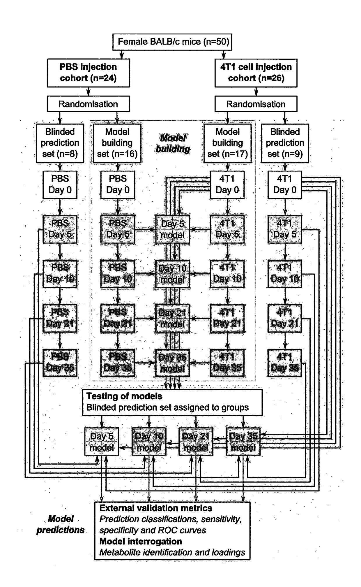
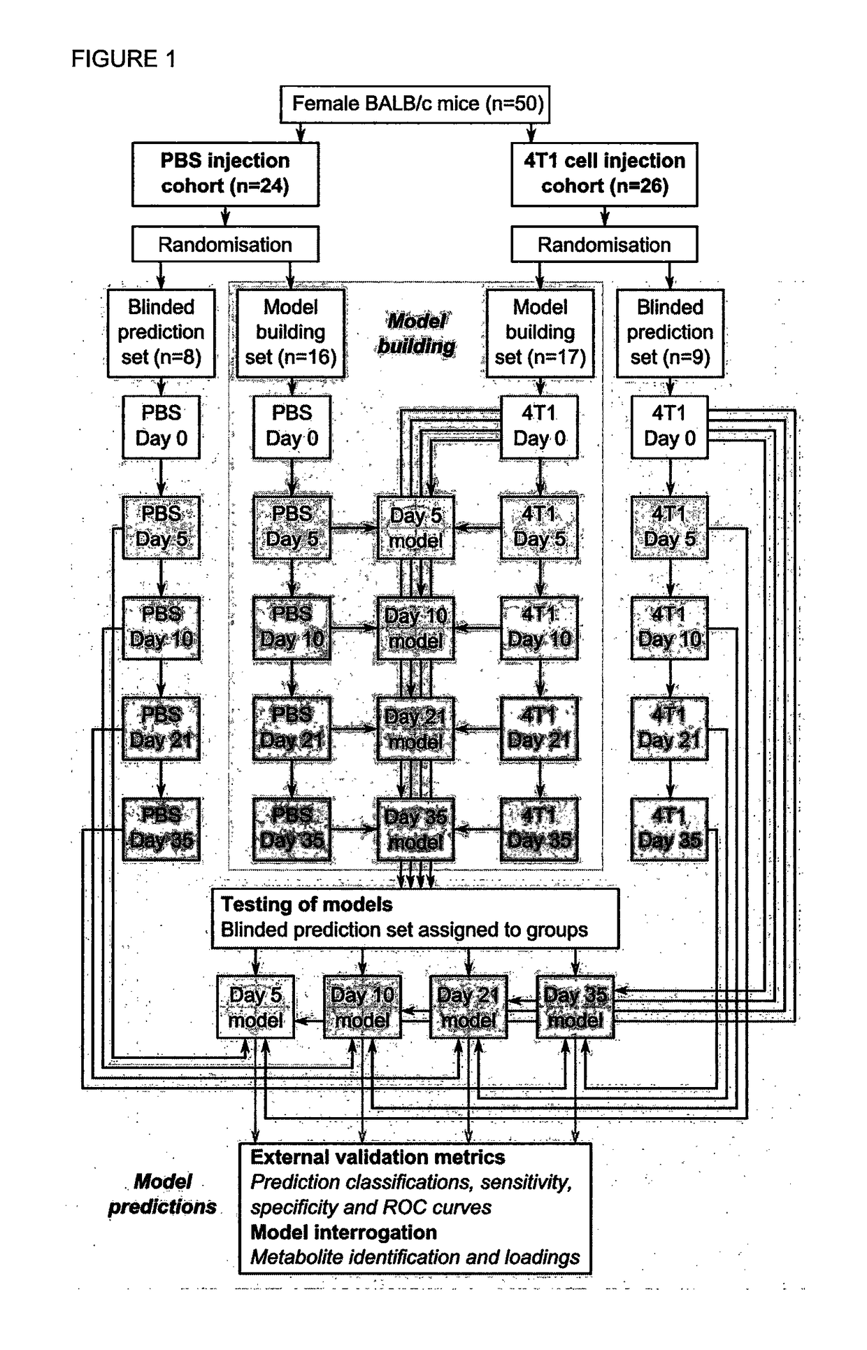
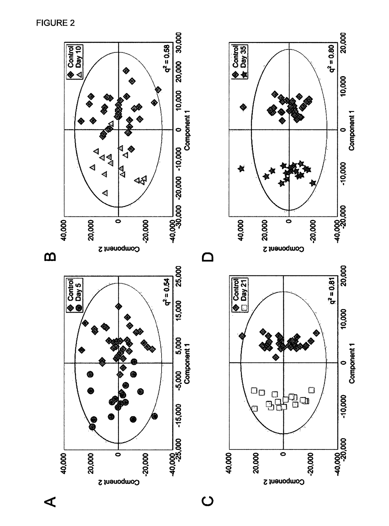
[0318]Four Orthogonal Partial Least Squares Discriminant Analysis (OPLS-DA) models were constructed separating NMR spectra of urine samples obtained from mice 5, 10, 21 or 35 days after 4T1-GFP cell injection from their respective control cohorts. The study design, including control groupings, is shown in FIG. 1. All four models were significantly predictive with models from later time points being stronger than those from earlier time points (FIG. 2). For days ...

example 2

[0327]In order to validate the proposed biofluid metabolomics approach for the early detection of brain metastasis, two further experimental models were considered: (i) metastatic human breast carcinoma cells (MDA-231-BR-GFP) injected intracerebrally in SCID mice; and (ii) metastatic mouse melanoma cells (B16F10) injected intracerebrally in syngeneic C57BL / 6 mice.

Methods

[0328]For each model, urine samples were collected from animals 10 days after intracerebral injection with tumour cells (MDA-231-BR-GFP, n=5; B16F10, n=6) or PBS-vehicle alone (n=6 and n=5, respectively). Samples from naïve animals (n=5 for each cell line) were also collected. Samples from naïve and PBS-injected animals were combined into a single control cohort, as for the 4T1-GFP models (see Example 1). In each case, OPLS-DA models were constructed and q2 values determined. Integral regions contributing strongly to each separation were identified.

Results

[0329]In each case, models with q2 values greater than the def...

example 3

[0332]In order to validate the repeatability of the intracerebrally injected 4T1-GFP-based models, a small set of animals was used to replicate the models. This second cohort of animals, hereafter referred to as Cohort B, was prepared by an independent researcher (AMD) and is thus distinct from the cohort used for the main study, hereafter referred to as Cohort A.

Methods

[0333]Cohort B included urine samples from female BALB / c mice collected 10 days after intracerebral injection of either 4T1-GFP cells (n=6) or PBS vehicle alone (n=5), as well as samples from age-matched naïve mice (n=5). The samples from the PBS-injected animals and the naïve animals were combined into a single control cohort, as described in the main text (n=10). Cohort B is independent to cohort A in four important ways: (i) the independent researcher performed all scientific steps including cell culture, animal handling, solution preparation and sample analysis; (ii) cohorts A and B were temporally separated by >...


PUM

PropertyMeasurementUnit
pHaaaaaaaaaa
pHaaaaaaaaaa
pHaaaaaaaaaa

Abstract

An in vitro method for diagnosing a brain tumour in a human test subject comprising: determining the concentration of at least two metabolites comprised in a biofluid sample obtained from the human test subject; comparing the concentration of the at least two metabolites in the biofluid sample with the concentration of the same at least two metabolites in at least one reference standard obtained from a non-tumour bearing subject and/or a tumour bearing subject; and identifying a concentration difference for each of the at least two metabolites in the biofluid sample relative to the reference standard; wherein the concentration difference for each of the at least two metabolites in the biofluid sample correlates with the presence of a brain tumour. The invention also relates to a data-storage medium comprising data obtained by a method of the invention.

Description

FIELD OF THE INVENTION[0001]The present invention relates to an in vitro method for diagnosing a brain tumour in a human test subject as well as a data-storage medium comprising data obtained by a method of the invention.BACKGROUND[0002]Metastasis, the spread of a tumour from its primary site to a distant location, is a leading cause of cancer mortality and remains a significant hurdle. Approximately 20-40% of cancer patients will develop brain metastases. Lung cancer, breast cancer and melanoma are the most common primary tumours in patients with brain metastasis and between them they account for 67 to 80% of all cases. Critically, brain metastases present a particularly tough clinical challenge owing to the nature of the blood-brain barrier (BBB) which thwarts attempts to diagnose tumours and also inhibits their treatment with most systemically active therapeutic agents.[0003]Brain metastases are diagnosed by imaging methods, for which the current clinical gold standard is gadolin...

Claims


Application Information

Patent Timeline
Patent Type & Authority Applications(United States)
IPC IPC(8): G01N33/574
CPCG01N33/57407
Inventor SIBSON, NICOLA R.ANTHONY, DANIEL C.DICKENS, ALEX M.LARKIN, JAMES R.
Owner OXFORD UNIV INNOVATION LTD
Why Eureka
Try Eureka