Mobile microscope

a microscope and mobile technology, applied in the field of optical microscopic imaging, can solve the problems of inability to perform early diagnosis and treatment, sparse network of permanent laboratories capable of performing the required tests, etc., and achieve the effect of convenient portability and small structur

Active Publication Date: 2018-08-30
UNIVERSITY OF HELSINKI
View PDF0 Cites 4 Cited by
  • Summary
  • Abstract
  • Description
  • Claims
  • Application Information

AI Technical Summary

Benefits of technology

[0007]The invention provides advantages. Due to sufficient optical capabilities of the at least one polymeric lens, the detector and the objective lens may be maintain

Problems solved by technology

Early diagnosis and treatment is at the present not possible in particular in developing countries, where traffic communicati

Method used


Image

  • Mobile microscope
  • Mobile microscope
  • Mobile microscope

Examples


implementation example

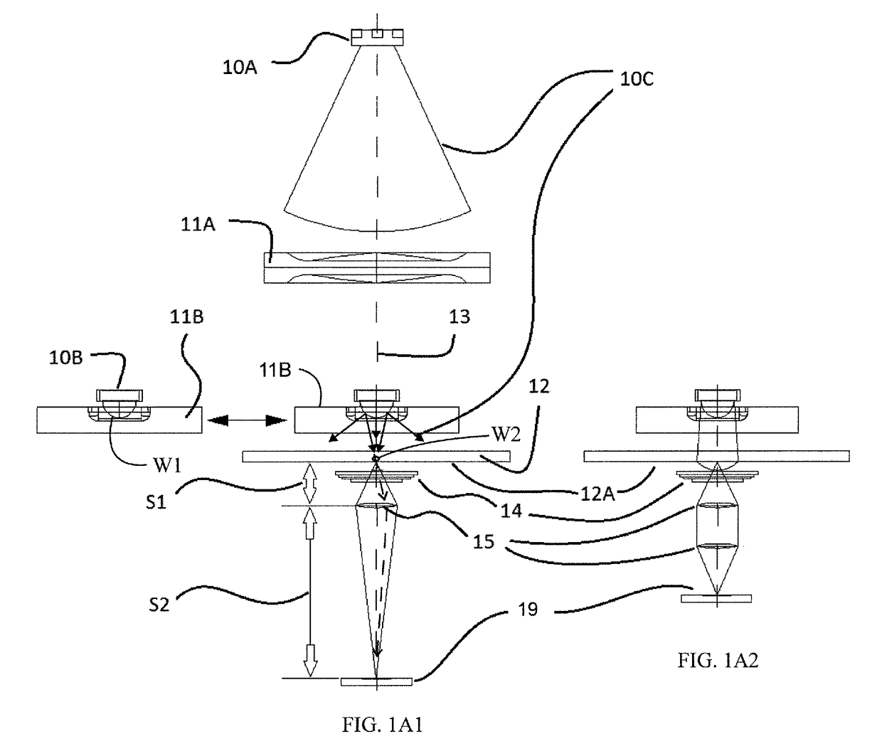
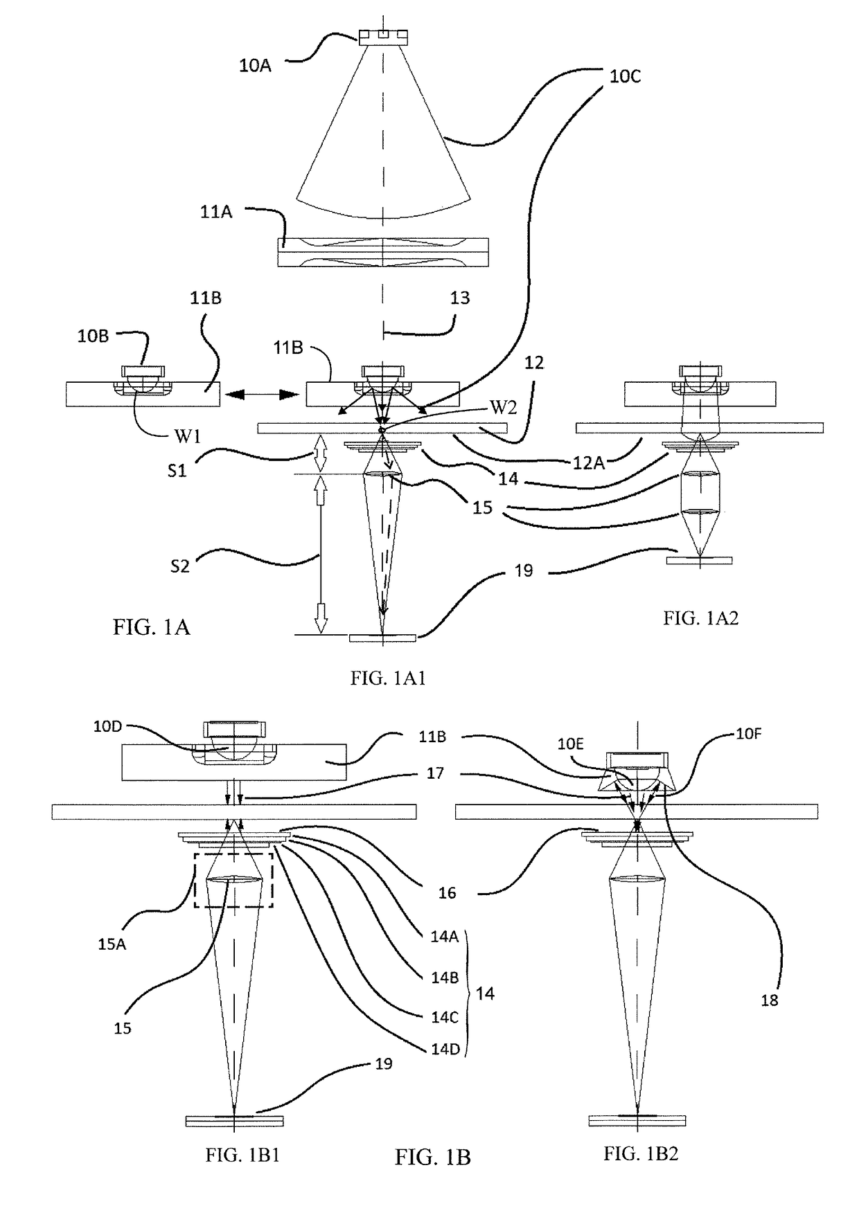
[0097]A mobile microscope may be built according to the design principles described above and shown in FIG. 1A, for example. At least one inexpensive plastic optical element used in mobile phone camera systems may be employed. The tube length of the microscope built may be decreased to about one tenth of a common microscope, for example, to about 16 mm, in order to reduce the size. Normal microscopes, using changeable objectives, which are used for forming different magnifications, yield captured image sizes from about 100 μm to 400 μm, whereas the microscope with the at least one plastic optical element may have a captured image size in the range of about 1000 μm, for example. The microscope may have a maximum fixed field of view of about 1.0×0.7 mm2 which is the imaged area W2. When using the maximum array size (2592×1944) the pixel size in the image may be about 400 nm.

[0098]The white light source may comprise a white LED, comprising blue emission LED, and adjacent polycrystallin...


PUM


Abstract

The invention relates to a mobile microscopic imaging device comprising a sample stage for holding a sample to be imaged, at least one light source for illumination of the sample, an imaging panel capable of capturing an image of the sample upon transmission illumination of the sample by the light source, and an optical magnification unit between the sample and the imaging panel for guiding light from the illuminated sample to the imaging panel so that a magnified image of at least portion of the sample is formed at the imaging panel. According to the invention, the optical magnification unit comprises a filter integrated polymeric lens assembly in a transmitted light fluorescence configuration which allows for both miniaturization of the device to a truly mobile level and reducing manufacturing costs.

Description

FIELD OF INVENTION[0001]The invention relates to optical microscopic imaging. In particular, the invention relates to a mobile microscope suitable for use in point-of-care diagnostics, for example.BACKGROUND ART[0002]Modern medical point-of-care (POC) methods benefit from the capability of high-quality optical diagnosis devices that are easily movable close to a patient to be diagnosed. The ability to capture high quality microscopic images of samples taken from patients without delay, for example for determining a disease, offers a possibility to provide rapid and accurate treatment for the patient. Early diagnosis and treatment is at the present not possible in particular in developing countries, where traffic communications / roads and vehicles are poor and the network of permanent laboratories capable of performing the required tests is sparse. However, there are many situations, also in developed countries, in which point-of-care would offer advantages for the patients or the car...

Claims


Application Information

Patent Timeline
IPC IPC(8): G02B21/00G02B21/26G02B21/16G02B5/20G02B5/22G02B5/28
CPCG02B21/0008G02B21/26G02B21/16G02B5/208G02B5/22G02B5/283
Inventor LUNDIN, JOHANSUUTALA, ANTTIMOILANEN, HANNU
Owner UNIVERSITY OF HELSINKI
Why Eureka
Try Eureka