Method of image support for a person carrying out a minimally invasive procedure with an instrument in a procedure site of a patient, x-ray apparatus, computer program and electronically readable data carrier

a technology for a patient and an instrument, which is applied in the field of image support for a person carrying out a minimally invasive procedure with an instrument in the procedure site of a patient, can solve the problems that the three-dimensional model data set only adequately describes the anatomy for a large number of applications, and achieves the effects of reducing the time of the minimally invasive procedure, high accuracy, and time-efficient and reliable monitoring

Active Publication Date: 2018-09-06
SIEMENS HEALTHCARE GMBH
View PDF3 Cites 4 Cited by
  • Summary
  • Abstract
  • Description
  • Claims
  • Application Information

AI Technical Summary

Benefits of technology

[0007]Methods of iterative reconstruction (often also called: algebraic reconstruction) of three-dimensional data sets from two-dimensional projection images are already largely known in the prior art. With iterative reconstruction methods of this kind, a three-dimensional model—the three-dimensional model data set—is ultimately calculated, which explains the recorded X-ray images (projection images). Different virtual comparison images are stimulated during the iterative reconstruction from the three-dimensional model data set to be determined and these are compared with the recorded X-ray images. The three-dimensional model data set is then iteratively optimized by minimizing the errors between the simulated comparison images and the recorded X-ray images. An iterative reconstruction method of this kind can also be carried out with a small number of X-ray images, in other words with sparsely occupied coverage in respect of the projection directions, wherein there is then an underdetermining, however, and the resulting three-dimensional model data set only inadequately describes the anatomy for a large number of applications.
[0011]To summarize, the invention therefore proposes associating the two-dimensional X-ray images recorded during a procedure in order to determine an inaccurate or incomplete three-dimensional model data set of the underlying anatomy in the procedure site, in which, in contrast to a sufficiently accurate, complete three-dimensional model data set, the expected image quality is sufficient only for simulated projection images, whose recording geometry corresponds to that of an acquired X-ray image considered in the iterative reconstruction. Supporting images can therefore be generated for the person without X-ray images having to be specially recorded. This enables a significant reduction in the time of the minimally invasive procedure as well as in the radiation dose administered, in particular if the position of a model object, in particular the medical instrument, actually acquired using X-rays can be acquired otherwise.
[0012]Use of the three-dimensional model data set has further advantages compared to an intended solution in which simply only the model object added by the further information is forward projected and superimposed in the X-ray images. Firstly, due to the fact that the iterative reconstruction is performed on the basis of a plurality of X-ray images, a smaller shift in the anatomy, which occurs, for example, due to inaccuracies in the determination of the recording geometries or small movements of the patient, between recording of these X-ray images is automatically compensated, resulting in a consistent overall image. Secondly, however, anatomical objects are, at least in principle, depicted in the three-dimensional model data set, so the arrangement relative to these anatomical objects is also given with the addition of an item of further information in the three-dimensional model data set, therefore masking effects can be correctly reproduced.
[0013]In a first advantageous, specific application of the present invention it can be provided that as a further item of information, a three-dimensional instrument model on the basis of a current item of position information of the instrument in the procedure site is inserted into the model data set and the projection of the instrument model into supporting images showing the recording geometries are determined and displayed. If, therefore, a three-dimensional item of position information of the instrument, which can of course also include an item of orientation information in addition to purely an item of location information, can be determined, a corresponding instrument model can be included as further information in the three-dimensional model data set with the current position and orientation. Instrument models of this kind can be easily derived, for example by means of dimensions and material properties of the medical instrument known from databases, which instrument can be, for example, a tool and / or an implant. Without recording X-ray images, or at least with a reduced number of X-ray images, the person is therefore provided with supporting images, which show the instrument at its current position relative to the anatomy in the procedure site. This enables highly accurate, time-efficient and reliable monitoring of the instrument positioning.
[0025]One development of the invention also provides that the model data set is updated when a current X-ray image is rerecorded in at least one of the recording geometries, in particular after 2D-3D registering of the model data set and the current X-ray image. In this way, inaccuracies due to sensors, patient movements and the like can be minimized in that 2D-3D registering takes place before new, current X-ray images are considered in the model data set.

Problems solved by technology

An iterative reconstruction method of this kind can also be carried out with a small number of X-ray images, in other words with sparsely occupied coverage in respect of the projection directions, wherein there is then an underdetermining, however, and the resulting three-dimensional model data set only inadequately describes the anatomy for a large number of applications.

Method used


Image

  • Method of image support for a person carrying out a minimally invasive procedure with an instrument in a procedure site of a patient, x-ray apparatus, computer program and electronically readable data carrier
  • Method of image support for a person carrying out a minimally invasive procedure with an instrument in a procedure site of a patient, x-ray apparatus, computer program and electronically readable data carrier
  • Method of image support for a person carrying out a minimally invasive procedure with an instrument in a procedure site of a patient, x-ray apparatus, computer program and electronically readable data carrier

Examples


Embodiment Construction

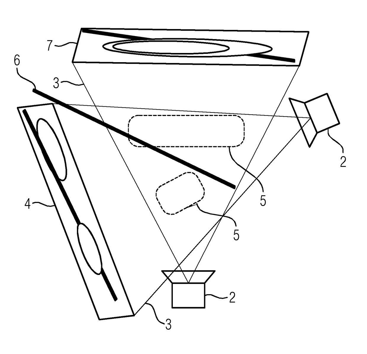
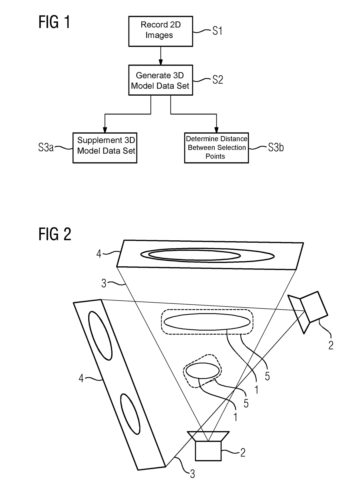
[0039]Referring now to the figures of the drawings in detail and first, particularly to FIG. 1 thereof, there is shown a basic flowchart of an exemplary embodiment of an inventive method. A person carrying out a minimally invasive procedure is to be supported by images. To enable this, it is firstly provided in a step S1 that by means of an X-ray apparatus, two-dimensional X-ray images are recorded using an X-ray apparatus in recording geometries chosen by the person and which correspond to different projection directions specific to the procedure. In the present case this is an X-ray apparatus having a C-arm, which can also be flexibly positioned at the procedure site in order to record the X-ray images. If an example from traumatology is used, the person can, for example, select the recording geometries specific to the procedure such that the correct position of an implant as the medical instrument can be optimally assessed. In the present case, by way of example two X-ray images,...


PUM


Abstract

In a method of image support for a person carrying out a minimally invasive procedure with an instrument in a procedure site of a patient, one two-dimensional X-ray image respectively of the procedure site is recorded by an X-ray apparatus in at least two recording geometries implementing different projection directions and chosen specific to the procedure, in particular by the person. A three-dimensional model data set of the procedure region is reconstructed by iterative reconstruction from the X-ray images. At least one two-dimensional supporting image corresponding to one of the recording geometries is determined by incorporating at least one item of further information by re-projection of the model data set supplemented by the further information and is displayed for the user.

Description

CROSS-REFERENCE TO RELATED APPLICATION[0001]This application claims the benefit, under 35 U.S.C. § 119, of German patent application DE 10 2017 203 438.6, filed Mar. 2, 2017; the prior application is herewith incorporated by reference in its entirety.BACKGROUND OF THE INVENTIONField of the Invention[0002]The invention relates to a method of image support for a person carrying out a minimally invasive procedure with an instrument in a procedure site of a patient, wherein one two-dimensional X-ray image respectively of the procedure site is recorded by an X-ray apparatus in at least two recording geometries implementing different projection directions and chosen specific to the procedure, in particular by the person. In addition, the invention relates to an X-ray apparatus, to a computer program and to an electronically readable data carrier.[0003]With minimally invasive surgical procedures, for example in traumatology, the appropriate medical instruments, for example tools and / or imp...

Claims


Application Information

Patent Timeline
Patent Type & Authority Applications(United States)
IPC IPC(8): A61B34/10G16H30/40G06T19/00G06T11/00
CPCA61B34/10G16H30/40G06T19/00G06T11/008A61B6/4441A61B6/466A61B2034/105A61B2034/102G06T2219/012G06T2219/008G06T2211/424A61B34/20A61B2034/2051A61B6/12A61B6/5205G06T11/006
Inventor GEMMEL, ALEXANDERKREHER, BJOERNKLEINSZIG, GERHARDWEITEN, MARKUS
Owner SIEMENS HEALTHCARE GMBH
Why Eureka
Try Eureka