A system and method for detection of suspicious tissue regions in an endoscopic procedure

a tissue region and endoscope technology, applied in image data processing, medical science, image enhancement, etc., can solve the problems of difficult detection, difficult detection of sessiles, difficult detection of polyps, etc., and achieve the effect of reducing the chances of finding and increasing the chances of finding polyps in images

Active Publication Date: 2018-09-06
MAGENTIQ EYE LTD
View PDF0 Cites 76 Cited by
  • Summary
  • Abstract
  • Description
  • Claims
  • Application Information

AI Technical Summary

Benefits of technology

[0054]In some embodiments, the method further comprises the step of identifying reflections in each frame or portion of frame and creating additional accompanying image in which each reflection is replaced with the surrounding intensity, colors and texture. Reflections increase the chances of finding a polyp in the image.
[0055]In some ...

Problems solved by technology

It is more difficult to detect small polyps (less than 6 mm) than large polyps, sessile are more difficult to detect than pedunculated since they are less salient form the colon tissue.
Flat polyps are more difficult to detect than bump polyps, and very small (less than 2 mm) flat polyps are usually the most difficult to detect.
In addition ...

Method used


Image

  • A system and method for detection of suspicious tissue regions in an endoscopic procedure
  • A system and method for detection of suspicious tissue regions in an endoscopic procedure
  • A system and method for detection of suspicious tissue regions in an endoscopic procedure

Examples


Embodiment Construction

[0088]In the following detailed description of various embodiments, reference is made to the accompanying drawings that form a part thereof, and in which are shown by way of illustration specific embodiments in which the invention may be practiced. It is understood that other embodiments may be utilized and structural changes may be made without departing from the scope of the present invention.

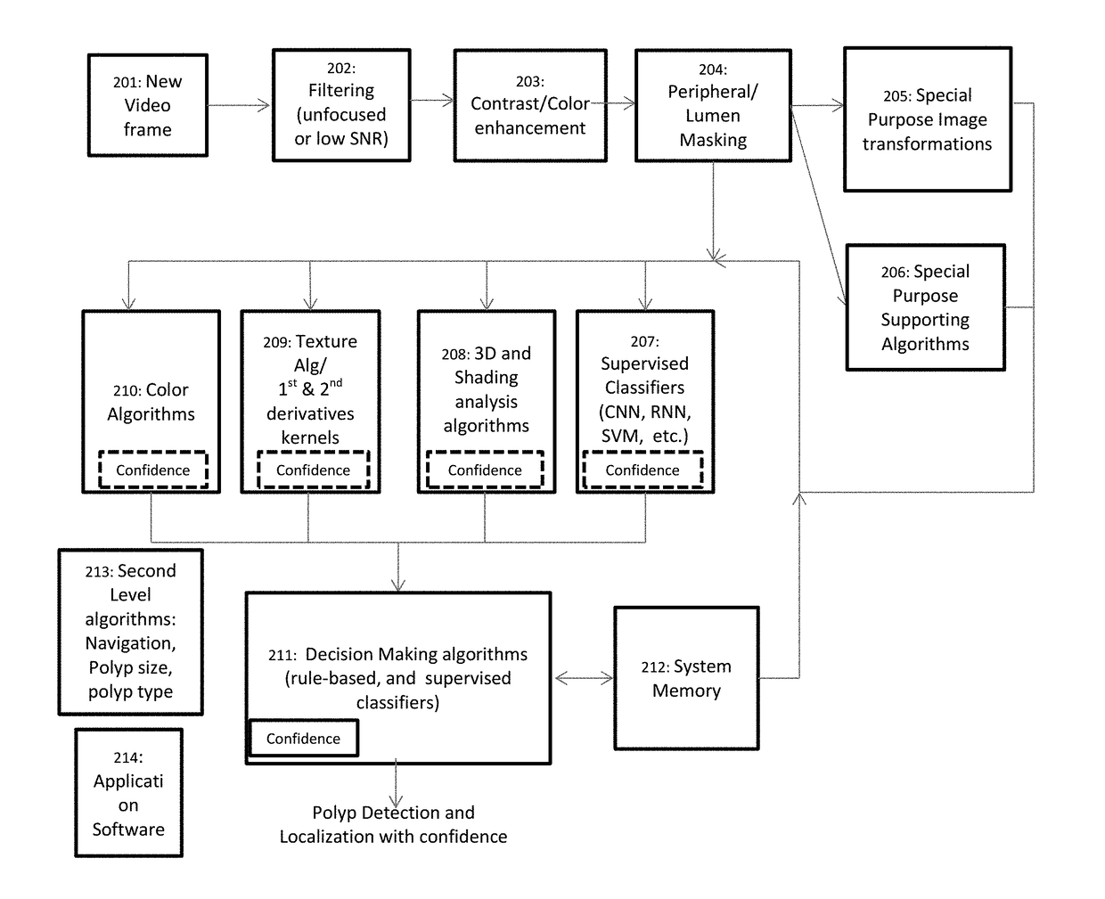
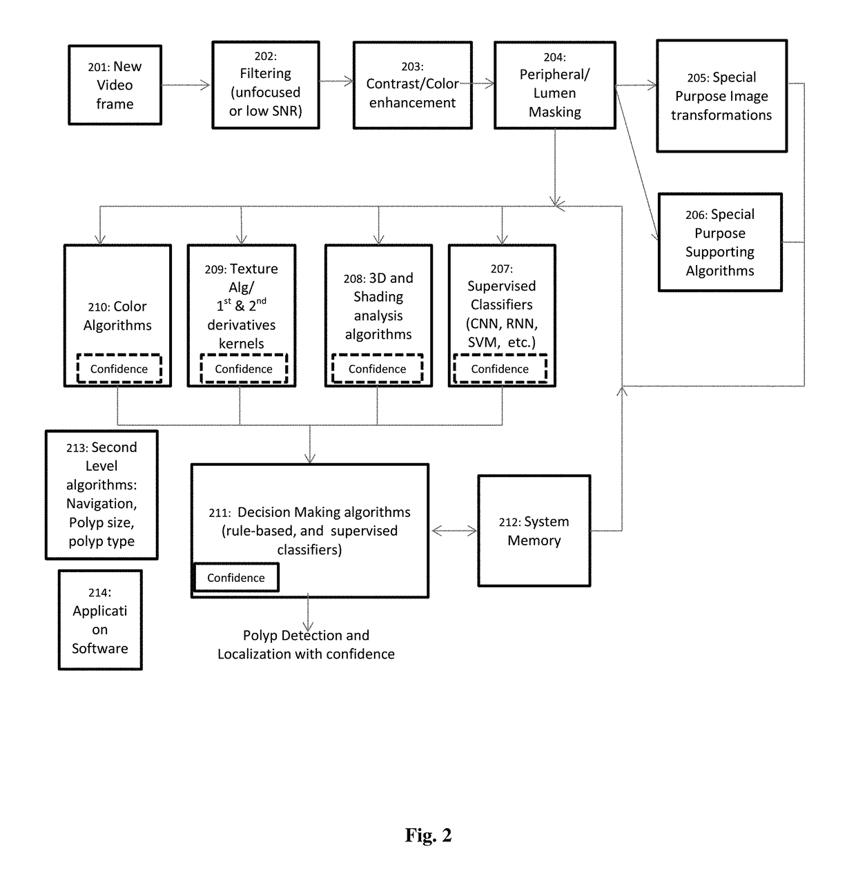
[0089]The system of the invention is adapted for automatically detecting the presence and location of suspicious tissue regions in endoscopic images. For clarity purposes, the disclosure details automatically detecting polyps in a colonoscopy procedure, though any person skilled in the art will immediately understand how to customize the system and method described to identify suspicious tissue regions in images of other endoscopic procedures in other cavities of the body such as a sigmoidoscopy examination, or any other endoscopic procedure.

[0090]The system and method of the invention examin...


PUM


Abstract

An image processing system connected to an endoscope and processing in real-time endoscopic images to identify suspicious tissues such as polyps or cancer. The system applies preprocessing tools to clean the received images and then applies in parallel a plurality of detectors both conventional detectors and models of supervised machine learning-based detectors. A post processing is also applied in order select the regions which are most probable to be suspicious among the detected regions. Frames identified as showing suspicious tissues can be marked on an output video display. Optionally, the size, type and boundaries of the suspected tissue can also be identified and marked.

Description

TECHNICAL FIELD[0001]The present invention relates to image processing in general and in particular to a system and method for detecting suspicious regions in an endoscopic procedure.BACKGROUND ART[0002]An endoscopic procedure means searching and screening inside the human body by means of a medical device (the endoscope) at a hollow organ or a cavity of the body for medical purposes. Unlike most other medical imaging devices, endoscopes are inserted directly into the organ, and usually use an optical camera in the visible frequencies, or near visible frequencies (such as infra-red beams) in order to produce images and video frames from within the organ. Usually, the endoscopic procedure comes to test whether there are suspicious local regions in the tested organ, such as polyps, tumor, or evidence of cancerous cells.[0003]A colonoscopy procedure is an endoscopic test of the large bowel (large intestine) and the distal part of the small bowel with an optical camera (usually with a C...

Claims


Application Information

Patent Timeline
IPC IPC(8): G06T7/00A61B1/31A61B1/00
CPCG06T7/0012A61B1/31A61B1/00009G06T2207/10068G06T2207/10016G06T2207/30096G06T2207/10028G06T2207/30028G06T7/246G06T2207/30032G06T2207/20084A61B1/000096A61B1/000094A61B1/000095
Inventor ZUR, DROR
Owner MAGENTIQ EYE LTD
Why Eureka
Try Eureka