Method and apparatus for vascular and visceral clipping

a technology of vascular and visceral wounds, applied in the field of trauma surgery, combat medicine, emergency medical services, etc., can solve the problems of increasing the chance of accelerated deterioration of the patient's condition, the optimized solution of sutures and current clips, and the inability to open visceral and vascular wound repair. , to achieve the effect of reducing the chance of tissue ingrowth, improving ease of removal, and low profil

Inactive Publication Date: 2008-01-29
DAMAGE CONTROL SURGICAL TECH
View PDF22 Cites 225 Cited by
  • Summary
  • Abstract
  • Description
  • Claims
  • Application Information

AI Technical Summary

Benefits of technology

[0010]The devices and methods described below provide for improved treatment of wounds, including achieving hemostasis and leakage control in hollow body vessels such as the small and large intestines, arteries and veins as well as ducts leading to the gall bladder and other organs. The devices are clips for hollow vessels, variously designed to make them suitable for emergency closure of vessels. The clip is capable of partially occluding a structure with a tangential wound or completely obstructing both ends of a completely divided blood vessel or hollow viscus. It is highly desirable that clamps be designed to be left behind for a period of time ranging from several minutes to several days without the attached handles. The clamps are designed to minimize the chance of tissue ingrowth, thus allowing for improved ease of removal. The ideal clamp would have a very low profile, secure holding properties, be incapable of eroding into other structures, would not crush or destroy the wall of the clamped vessel, even if left for a period of days, would be easy to apply, would evenly distribute the compression force on the vessel, would be able to straddle, in a partially occluding way, a tangential wound of a major vessel or bowel. Features of the vascular and bowel clips include broad, controlled, force distribution on the tissue, even force distribution, both longitudinally and laterally on the vessel or bowel tissue. Additional key features of the vascular and bowel clips of the present invention include controlled movement, ease of placement, ease of locking in place, ease of removal, biocompatibility for medium to long-term implantation, minimal projections away from the clip, lack of sharp edges to cause further trauma during placement, and removal of the clip applier so that there are no long surfaces projecting from the clip area. The vascular and bowel clips may be placed through an open surgical access site or through a laparoscopic access and manipulation system.
[0011]Once a clip has been placed, it remains in place either temporarily or permanently. Temporary placement necessitates removal of the clip after a period of time. Long-term placement necessitates that the clip be able to sustain its function indefinitely. In this application, the clip is fabricated from materials that permit medium to long-term implantation. The clip design minimizes undercuts and features that would promote tissue ingrowth, thus restricting removal. In another embodiment, the clip may be fabricated, completely or partially, from resorbable materials that obviate the need to remove the clip in a subsequent surgical procedure. The clip applier is fabricated from materials that are suitable for short-term tissue or vascular contact. The clips themselves are fabricated from materials with smooth outer surfaces that do not encourage tissue or clot ingrowth. Thus, the clips may be removed with minimal re-bleeding.
[0012]The clips may be partially or entirely radiopaque so that they can be visualized on fluoroscopy or X-ray and easily located on subsequent follow-up.
[0013]The current medical practice of using sutures and current clips is not an optimized solution to open visceral and vascular wound repair. The current techniques almost always cause a tourniquet effect and require substantially more time to place than is desired, thus increasing the chance of accelerated deterioration of the patient's condition. The devices and methods described herein distinguish over the current medical practice because the present invention is tailored to the needs of open vascular or bowel repair. The clips have soft serrated jaws to grab the vessel wall and prevent spillage, but not strangulate the vasculature within the wall. They have short stubby grasping handles that are activated by tools that provide mechanical advantage and extension of reach into small spaces. The clips are suited for either open surgical implantation and removal, or they are suited for laparoscopic placement and removal using specialized access, grasping and delivery instruments. When the clips of the present invention are removed from the patient, re-bleeding does not occur because there is minimal penetration of the wound tissues or clot into the interstices of the clips.
[0014]Another feature of the clips is reduction in the length of those portions of the implantable clip (the actuating lever arms) that projects away from the actual clamping surface. This minimization of projection away from the clamping surface is accomplished by use of more than one hinge point, telescoping members and applier grasping points located between the hinge point and the clamping surface. In another embodiment, the grasping tabs projecting away from the clip are minimized by folding them in a direction opposite to that in which they are compressed to open the clip. Folding the tabs back onto the clip allows the tabs to be completely or nearly completely eliminated as a projection. The folding tabs have their own hinge points or they rotate around the main clamp axis. In another embodiment, the tabs are rotated perpendicular to the direction of compression to move them out of the way so that they do not project away from the clip jaws.

Problems solved by technology

The current medical practice of using sutures and current clips is not an optimized solution to open visceral and vascular wound repair.
The current techniques almost always cause a tourniquet effect and require substantially more time to place than is desired, thus increasing the chance of accelerated deterioration of the patient's condition.

Method used


Image

  • Method and apparatus for vascular and visceral clipping
  • Method and apparatus for vascular and visceral clipping
  • Method and apparatus for vascular and visceral clipping

Examples


Embodiment Construction

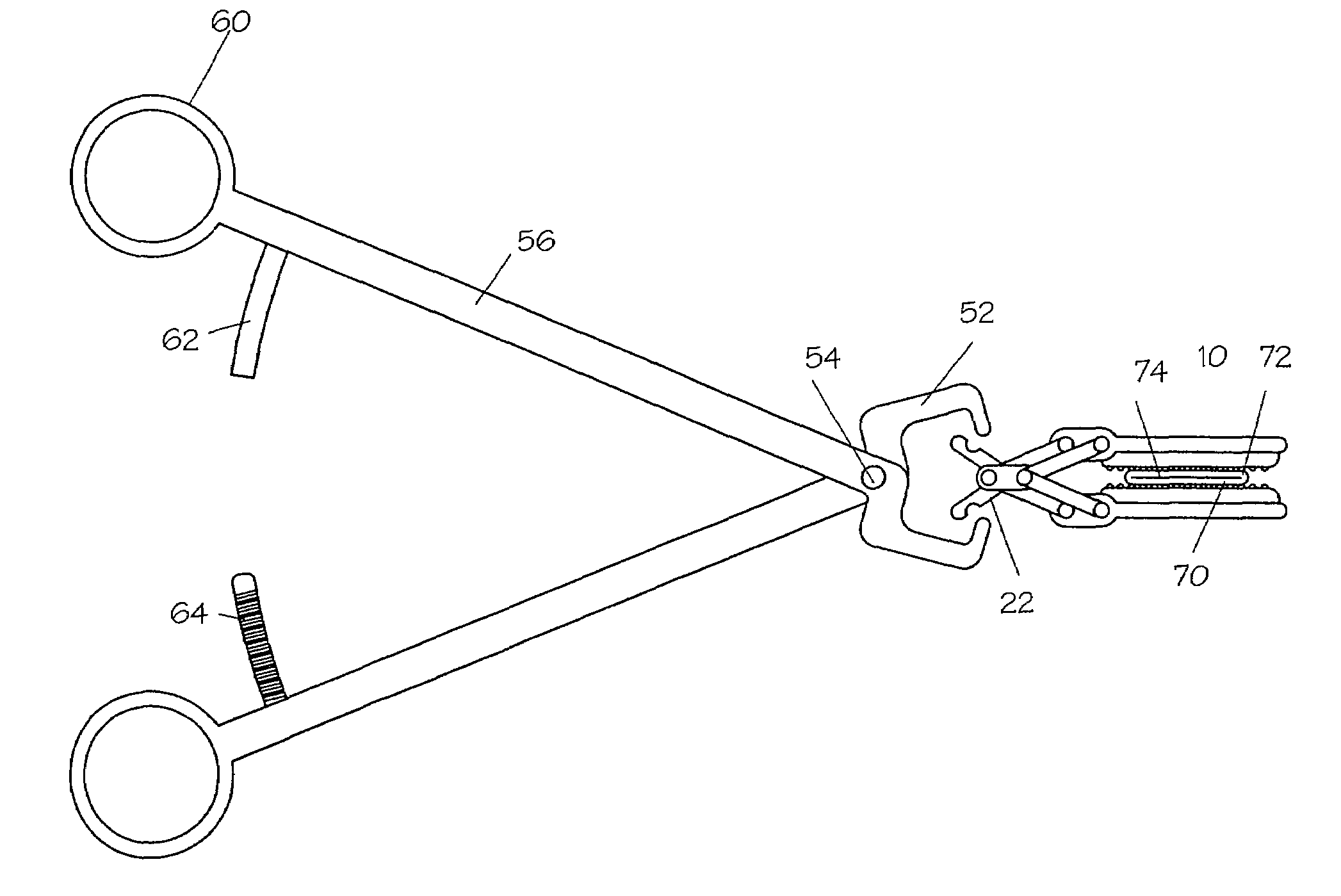
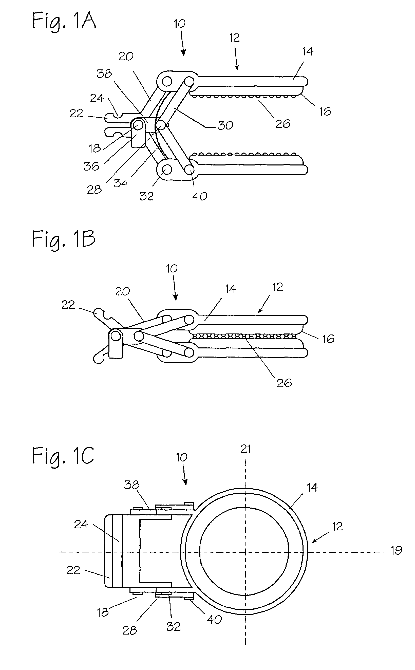
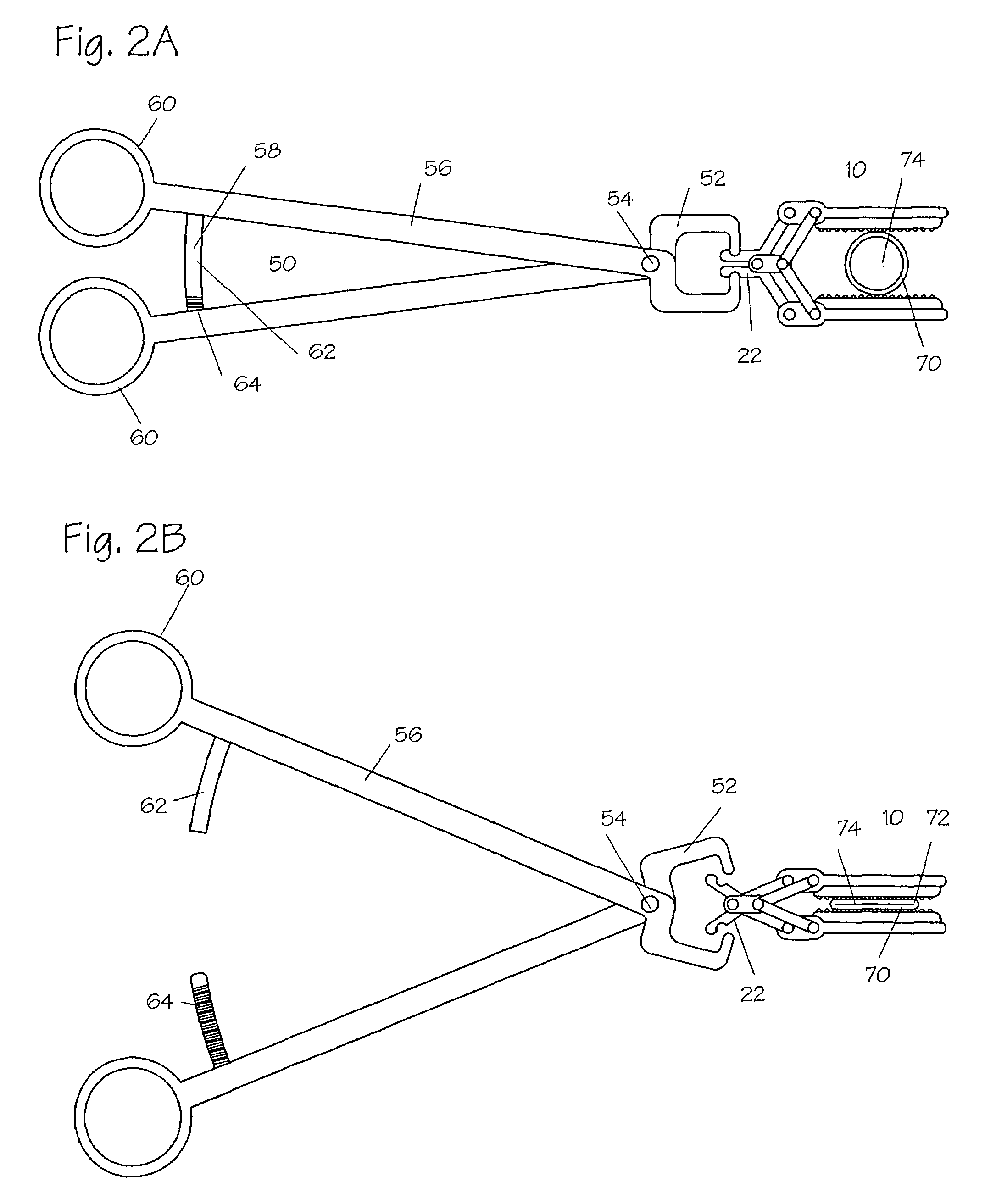
[0052]FIG. 1A illustrates a side view of a clip 10 of the present invention. The clip 10 comprises a plurality of jaws 12, further comprising a frame 14 and a pad 16, a main hinge 18, a plurality of main struts 20, a plurality of opening tabs 22, a plurality of grasping detents 24, and a plurality of optional serrations 26 on one or more of the pads 16. The clip 10 further comprises an optional secondary hinge 28, a plurality of optional secondary struts 30, a plurality of main pivot points 32, an optional spring 34, an optional lock 36, an optional hinge bracket 38, and a plurality of optional secondary pivot points 40.

[0053]Referring to FIG. 1A, the jaws 12 of the clip 10 are shown in their open configuration. The frame 14 provides rigid support and orientation for the pads.16. The top and bottom frames 14 are connected to a main hinge 18 through a plurality of main struts 20. Each of the main struts 20 is rigidly affixed to the opening tab 22 with the grasping detent 24 formed in...


PUM


Abstract

Devices and methods for achieving hemostasis and leakage control in hollow body vessels such as the small and large intestines, arteries and veins as well as ducts leading to the gall bladder and other organs. The devices and methods disclosed herein are especially useful in the emergency, trauma surgery or military setting, and most especially during damage control procedures. In such cases, the patient may have received trauma to the abdomen, extremities, neck or thoracic region. The devices utilize removable or permanently implanted, broad, soft, parallel jaw clips with minimal projections to maintain vessel contents without damage to the tissue comprising the vessel. These clips are applied using either standard instruments or custom devices that are subsequently removed leaving the clips implanted, on a temporary or permanent basis, to provide for hemostasis or leakage prevention, or both. These clips overcome the limitations of clips and sutures that are currently used for the same purposes. The clips come in a variety of shapes and sizes. The clips may be placed and removed by open surgery or laparoscopic access.

Description

[0001]This application claims priority benefit under 35 USC § 119(e) from U.S. Provisional Application No. 60 / 447,110 filed Jun. 9, 2003, and U.S. Provisional Application No. 60 / 410,635 filed Sep. 13, 2002.FIELD OF THE INVENTION[0002]The field of this invention is trauma surgery, combat medicine, and emergency medical services.BACKGROUND OF THE INVENTION[0003]As recently as the early 1990s, surgical operations for trauma were directed at the anatomic repair of all injuries at time of the initial operation. It was observed during these exercises that many patients became hypothermic, acidotic, and coagulopathic. Patients showing these three signs often died. Death often occurred in the operating room due to exsanguination, or postoperatively, due to the complications of prolonged shock and massive transfusion to replace blood lost as a result of the trauma.[0004]One of the most notable developments in the recent evolution of surgery has been the reintroduction of the concept of stage...

Claims


Application Information

Patent Timeline
Patent Type & Authority Patents(United States)
IPC IPC(8): A61B17/122A61BA61B17/08A61B17/10A61B17/12A61B17/128
CPCA61B17/08A61B17/10A61B17/122A61B17/128A61B17/083Y10T24/44376A61B2017/12004A61B17/1227
Inventor BUCKMAN, ROBERT F.LENKER, JAY A.KOLEHMAINEN, DONALD J.
Owner DAMAGE CONTROL SURGICAL TECH
Why Eureka
Try Eureka