Novel slice cultures and methods for diagnosing neuronal degeneration diseases

a neuronal degeneration and slice culture technology, applied in the field of slice cultures, can solve the problems of impaired neuronal function, neuronal damage and loss, and inability to allow free exchange of solutes, and achieve the effect of reducing the activity of ad biomarkers

Inactive Publication Date: 2018-10-11
ROWAN UNIVERSITY
View PDF0 Cites 0 Cited by
  • Summary
  • Abstract
  • Description
  • Claims
  • Application Information

AI Technical Summary

Benefits of technology

[0014]In another aspect, the present invention provides a method of evaluating the therapeutic effect of a compound comprising: contacting a test compound with a test slice culture, wherein said slice culture is treated with an agent before, after, or at the same time of contacting with the test compound, said agent induces one or more biomarkers of a neuronal degeneration disease, and said slice culture is selected from an organotypic brain slice culture and a retinal slice culture; measuring one or more biomarkers of a neuronal degeneration disease in said test slice culture; comparing the measurement of said biomarkers with a control or a baseline level to evaluate the therapeutic effect of the compound for treating or preventing the neuronal degeneration disease. The method of the present invention is applicable to evaluating compounds useful in managing various diseases including for example ALS, PD, AD and HD. The biomarkers may include Immunoglobulin G (IgG), cytoskeletal proteins such as GFAP, MAP2 and vimentin, calcium binding proteins such as S100B and visinin-like proteins, proteins that impact the cyclic GMP pathway such as membrane guanylate cyclases and their modulators In at least one embodiment, agents inhibiting or reducing the activity of the AD biomarkers may be used as therapeutic agents to treat AD, including Early-Stage AD.

Problems solved by technology

The endothelial cells of the BBB differ from those in other parts of the mammalian body in that they lack fenestrations and therefore do not allow for free exchange of solutes between the blood and the brain parenchyma.
Access of the previously excluded and potentially damaging blood-borne plasma elements to the brain interstitium, results in disruption of brain homeostasis, impaired neuronal function, and eventually, neuronal damage and loss.

Method used


Image

  • Novel slice cultures and methods for diagnosing neuronal degeneration diseases
  • Novel slice cultures and methods for diagnosing neuronal degeneration diseases
  • Novel slice cultures and methods for diagnosing neuronal degeneration diseases

Examples


example 1

Experimental Procedures
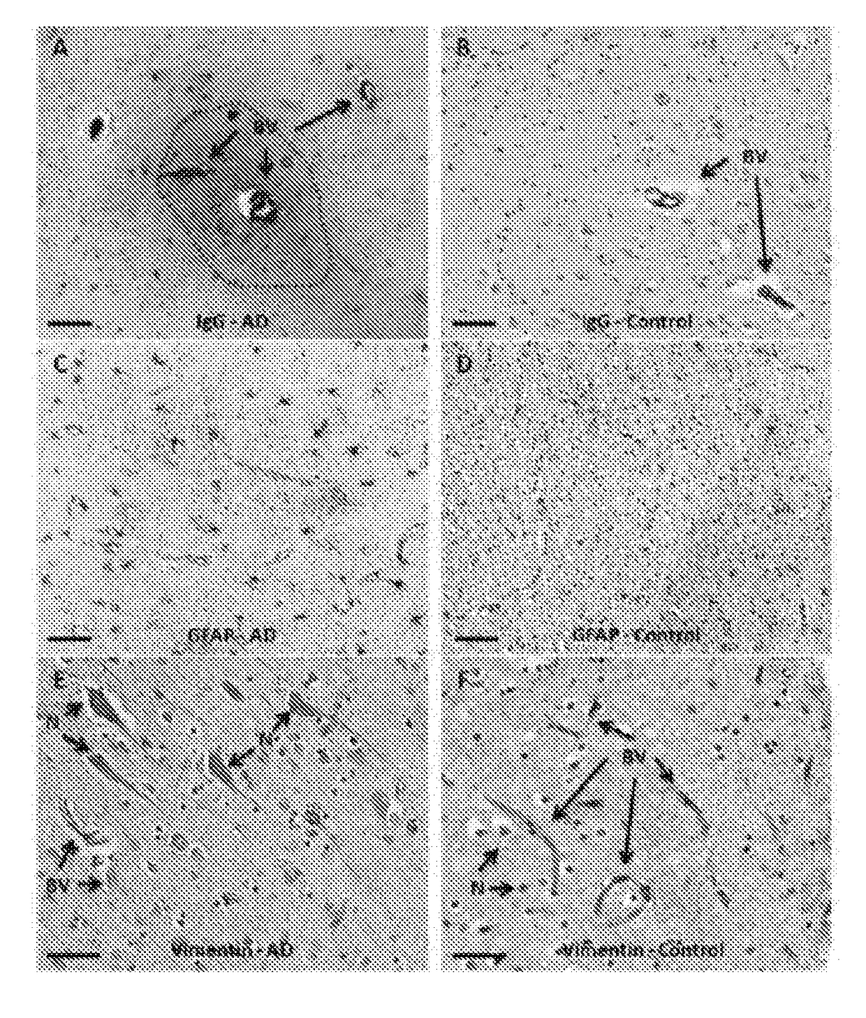
Antibodies

[0084]Human IgG antibodies (polyclonal, Cat. No. BA-3000, dilution=1:2000) and mouse IgG antibodies (polyclonal, Cat. No. BA-9200, dilution=1:2000) were obtained from Vectastain (Foster City, Calif.). GFAP antibodies were obtained from Millipore (Billerica, Mass.) (polyclonal, Cat. No. AB5804, dilution=1:1000). Vimentin antibodies were obtained from Sigma (Saint Louis, Mo.) (monoclonal, Cat. No. V6630, dilution=1:200). The specificity of each of these antibodies was confirmed via western blot or ELISA (data not shown).

Animals

[0085]C57BL / 6J mice were obtained from Jackson Laboratories (Bar Harbor, Me.) and used at 9 months of age. Mice were maintained on ad libitum food and water with 12-hour light / dark cycle in an AAALAC-accredited vivarium under a UMDNJ IACUC-approved protocol.

Primary Mouse Brain Organotypic Cultures

[0086]Primary mouse brain organotypic cultures were prepared as described previously ((Levin et al., Brain Res. 1298, 194-207, 2009). B...

example 2

[0106]Retinal slice cultures treated with histamine also display pathologies consistent with AD. As shown in FIG. 7-9, retina with BRB breakdown and expression of GFAP and MAP2 have been observed after treatment of retinal slice cultures with histamine.

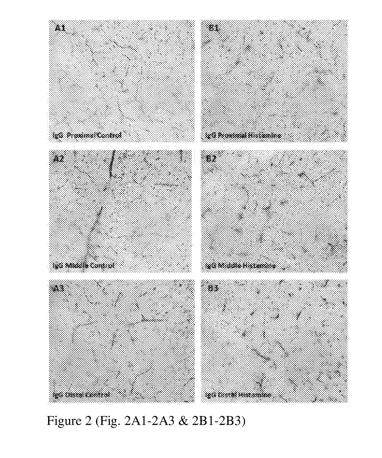
[0107]As shown in FIG. 7, retina with BRB breakdown and extravasated IgG surrounding blood vessels (indicated by dotted circles) are observed after histamine treatment (Panel: Hist). In untreated retinal slices, IgG is confined to BV lumen (Panel: Ctrl).

[0108]Histamine treatment also increases the expression of GFAP in retinal slices. FIG. 8 illustrates retinal slice cultures after treated for indicated time with histamine (0, 30, 60 or 90 min). The slices were then processed to generate cryosections for immunostaining. The expression of GFAP was monitored by immunostaining. GFAP is observed in green. The results are presented in two rows: the top one without the nuclei and the bottom one—with the nuclei in blue. A structural response...

example 3

[0110]The slice cultures of the present invention can also be used for evaluating or identifying compounds with therapeutic potential for treating or preventing neuronal degeneration diseases such as AD. As shown in FIG. 10, retinal slices were treated with histamine with or without lipoxin A4. Untreated samples served as control. The slices were then processed to generate cryosections for immunostaining. The location of IgG (an indicator of BRB breach) was monitored by immunostaining. IgG was observed in red. The results were presented in two rows: the top one without LXA4 and the bottom one—with LXA4 treatment. In the absence of LXA4, neurons were loaded with IgG and appeared in red (indicated by arrowheads) after histamine treatment. Addition of LXA4, however, conferred protection from these effects.


PUM

PropertyMeasurementUnit
timeaaaaaaaaaa
timeaaaaaaaaaa
timeaaaaaaaaaa

Abstract

The present invention relates novel slice culture systems which provide a quick, simple, and effective tool for investigating pathological changes associated with AD. Also provided are methods of diagnosing AD and identifying potentially therapeutic compounds.

Description

CROSS-REFERENCE TO RELATED APPLICATIONS[0001]The present application claims priority under 35 U.S.C. § 119(e) of U.S. Provisional Application No. 62 / 151,698, filed Apr. 23, 2015, the disclosure of which is hereby incorporated by reference in its entirety.FIELD OF THE INVENTION[0002]The present invention relates to slice cultures for studying various neuronal degeneration diseases and for developing novel approaches to diagnosing and treating such diseases.BACKGROUND[0003]Alzheimer's disease (AD) is a progressive neurological disease, mainly of the elderly, that is hallmarked by cognitive decline which results in loss of language and communication skills, difficulty in learning, loss of memory, and alterations in personality and mood. AD is the most common form of dementia, currently affecting over 5.5 million people in the United States and more than 35 million people worldwide. The pathological changes seen in AD include synaptic loss, dendrite retraction, neuronal cell death, infl...

Claims


Application Information

Patent Timeline
Patent Type & Authority Applications(United States)
IPC IPC(8): G01N33/50
CPCG01N33/5082G01N33/5091G01N2800/2821G01N2800/2835G01N2800/2857G01N2800/28G01N33/5008
Inventor VENKATARAMAN, VENKATNAGELE, ROBERT G.WU, HAO
Owner ROWAN UNIVERSITY
Why Eureka
Try Eureka