

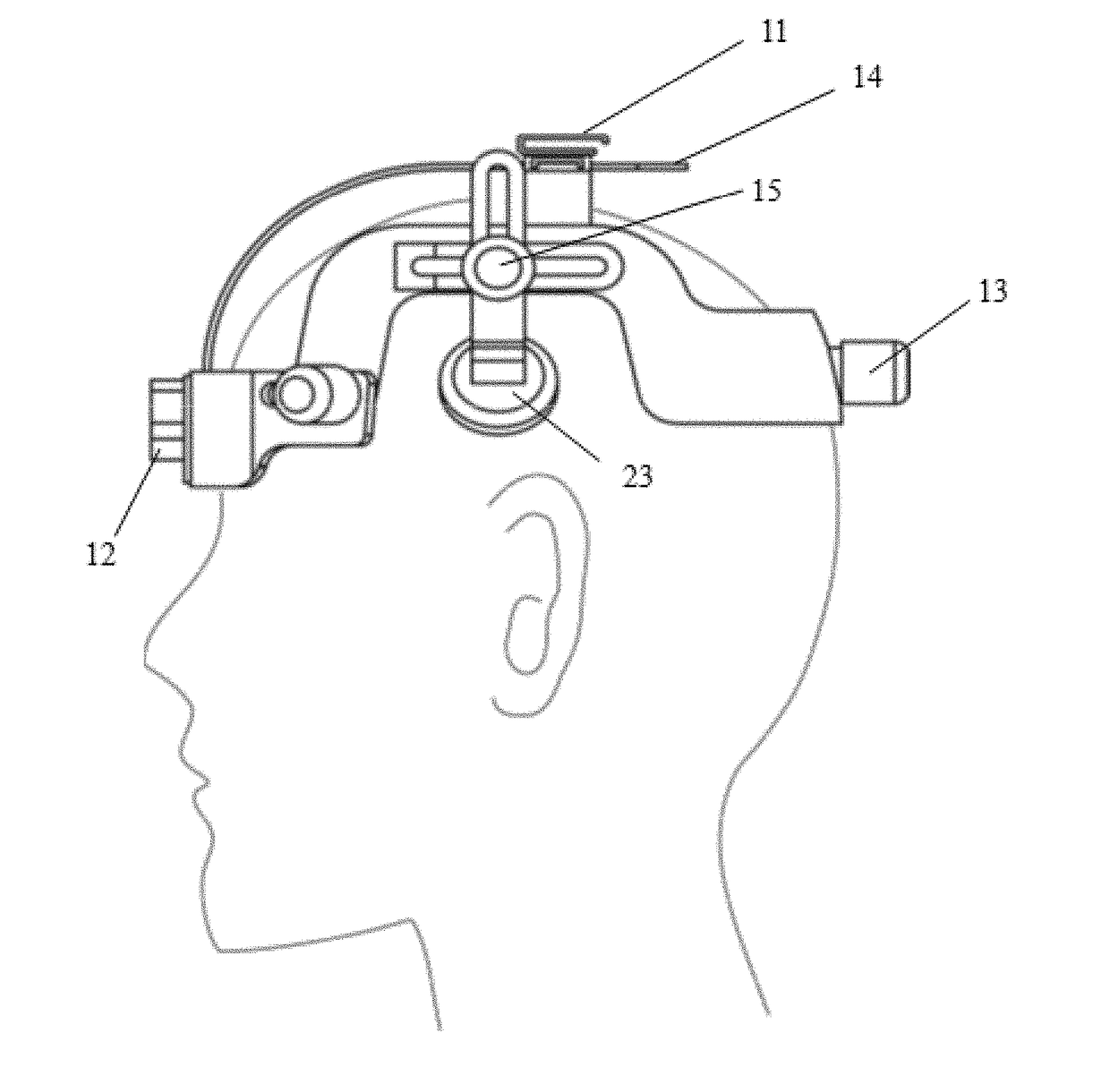
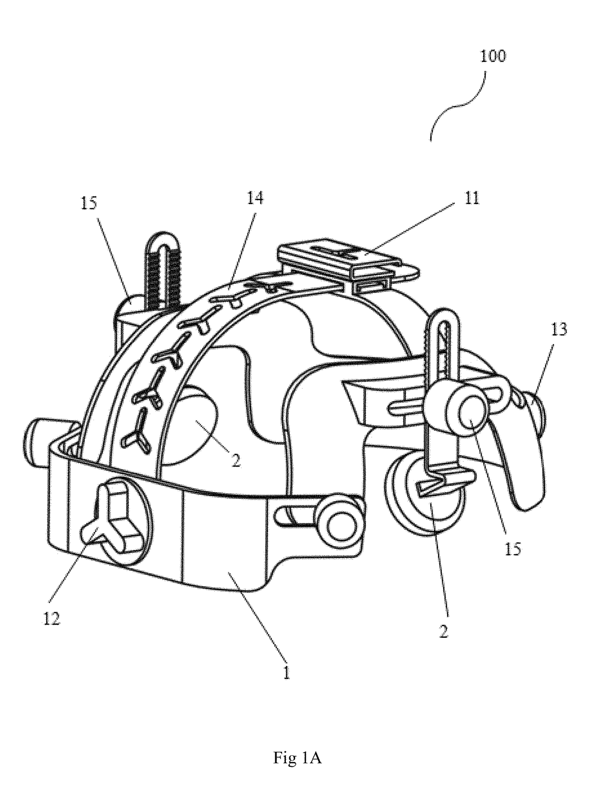
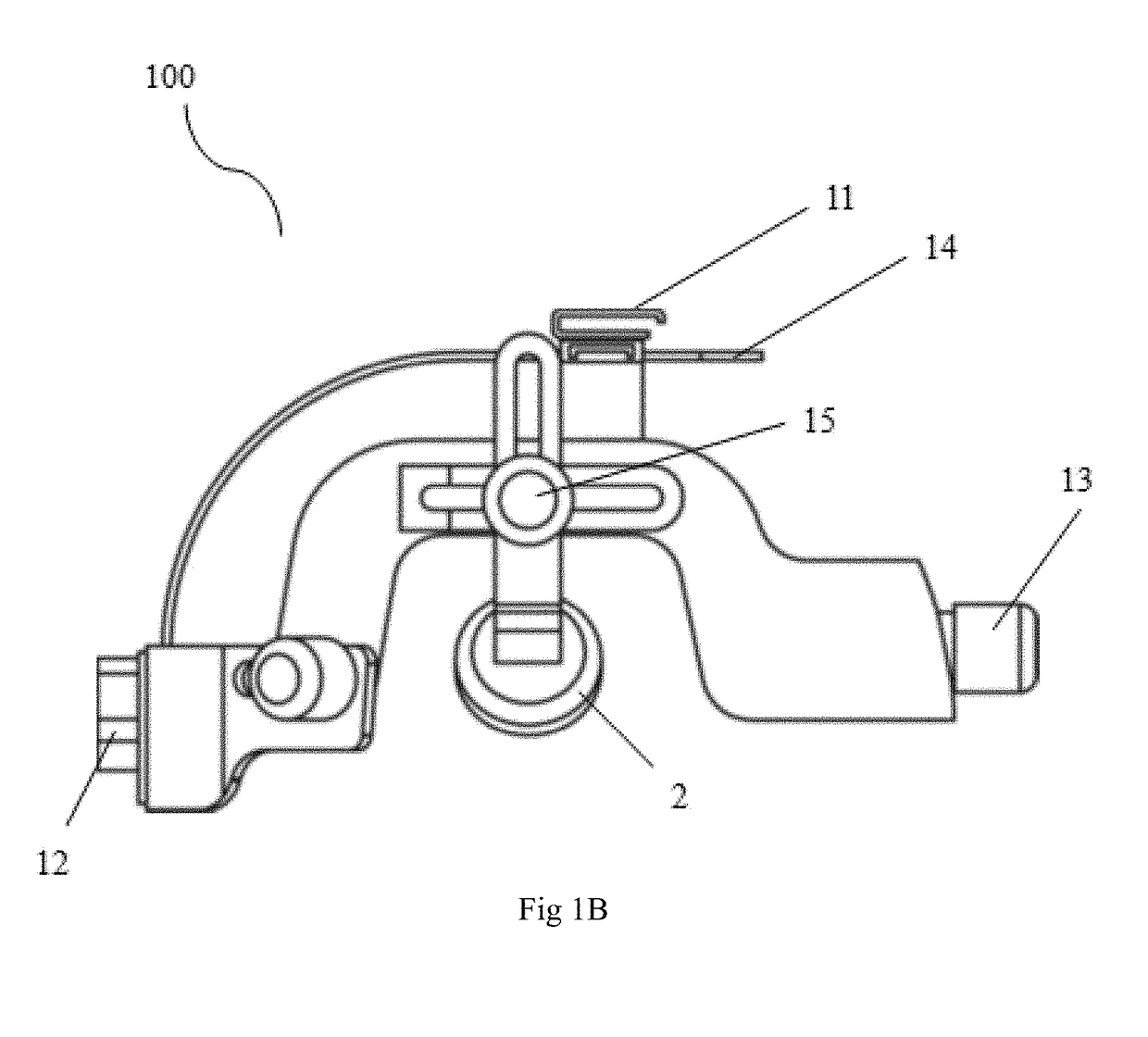
Ultrasound stimulation helmet
a technology of ultrasonic stimulation and helmet, which is applied in the field of ultrasonic stimulation helmet, can solve the problems of damage to the blood-brain barrier or the brain tissue, inability to completely cure neurodegenerative diseases, and current therapeutic drugs used only to control or slow down symptoms, etc., and achieve the effect of increasing or decreasing the expression level
- Summary
- Abstract
- Description
- Claims
- Application Information
AI Technical Summary
Benefits of technology
Problems solved by technology
Method used
Image

Examples
example 1
The Effect of Ultrasound Stimulation on the Endogenous Neurotrophic Factor in Brain
[0048]In the experimental group, the left and right hippocampus of Sprague-Dawley (SD) male rat were treated with low-intensity pulsed ultrasound stimulation with an output frequency of 0.3-1.0 MHz and an output power (ISPTA) of 10-720 mW / cm2 for seven consecutive days. The time of application was 15 minutes. Seven days later, SD male mice were sacrificed. Then detect the expression levels of the BDNF, GDNF and VEGF biomarker in brain of SD male rats were observed by Western blotting. The results are shown in FIGS. 4A, 4B and 4C, wherein the control group was the right and left hippocampi of SD male mice without applying low intensity pulsed ultrasound stimulation.
[0049]From the experimental results, the neurons of SD male mice (experimental group) stimulated with low intensity pulsed ultrasound stimulation could activate the expression of brain neurotrophic factors, comparing to the control group, in...
example 2
The Effect of Ultrasound Stimulation on Intracellular Calcium Concentration
[0051]Low-intensity pulsed ultrasound stimulation with an output frequency of 0.3-1.0 MHz and an output power (ISPTA) of 10-720 mW / cm2 was applied to rat brain astrocyte cells (CTX TNA2) several times (in the experimental group). The time of application was 5 minutes. The intracellular calcium concentration was measured by Western blotting at 0, 30, 60 and 120 seconds after the administration (in the control group) and before administration.
[0052]The results were shown in FIG. 6. Compared to the control group, the intracellular calcium concentration in the experimental group increased according to the increase of the ultrasound stimulation application time.
example 3
The Effect of Ultrasound Stimulation on TrkB phosphorylation in the Cell
[0053]Low-intensity pulsed ultrasound with an output frequency of 0.3-1.0 MHz and an output power (ISPTA) of 10-720 mW / cm2 was applied to rat brain astrocyte cells (CTX TNA2) several times (in the experimental group). The time of application was 5 minutes. The intracellular calcium concentration was measured by Western blotting at 0.5, 1, 2, and 4 hours after the administration (in the control group) and before administration.
[0054]The results were shown in FIG. 7, the level of intracellular phosphorylated TrkB in the experimental group was higher than the control group, and level of phosphorylated TrkB (p-TrkB) at 4 hours after ultrasound stimulation was the highest.
PUM
 Abstract
Description
Claims
Application Information