
Pharyngeal-enteric tube combination

a technology of pharyngeal and gastrointestinal tract, applied in the field of pharyngeal-enteric tube combination, can solve the problems of laceration of mucous membranes, ongoing daily material cost, and inability to reliably provide supplemental oxygen, so as to facilitate a secretion-free environment, improve patient autonomy, and improve measurable outcome

Inactive Publication Date: 2018-11-01
CEDARS SINAI MEDICAL CENT
View PDF0 Cites 1 Cited by
  • Summary
  • Abstract
  • Description
  • Claims
  • Application Information

AI Technical Summary

Benefits of technology

The invention disclosed in this patent provides a new method and device for reducing aspiration events, improving oropharyngeal secretion removal, and providing supplemental oxygen directly to the patient's larynx. The device is a combination of a suction catheter and a feeding tube, which conveniently reduces the amount, rate, and frequency of aspirations. Additionally, the device allows for the safe and reliable supply of supplemental oxygen to the patient's larynx, and results in measurable improvements in patient outcomes including reduction of hospital complications and increased patient comfort and autonomy. The method is convenient and predictable, and reduces the need for cumbersome oxygen delivery devices.

Problems solved by technology

However, intermittent oral suctioning: (1) requires, in the majority of patients, a vigilant caregiver day and night; (2) produces uncomfortable arousal each time a catheter is inserted into the throat; (3) elicits very frequently cough, gag, swallowing and apneic (breath holding) reflexes which are not only unpleasant experiences but also unwanted in patients with acute brain injury or healing wounds post-surgery, among many others; (4) can lead to bleeding and laceration of mucous membranes; and (5) requires a new, clean suction device replacement set at least every 24 hours resulting in ongoing daily material costs.
Nasal prongs (1) often dislodge from the nostrils which is commonly neither noted by the patient nor the care taker exposing the patient to unwitnessed hypoxemic episodes; (2) are frequently irritating as they reside and jiggle within the highly sensitive mucosa of the nostril cavity; and (3) do not reliably provide supplemental oxygen as many patients are dominant mouth-breathers, especially during sleep.
Face masks and tends are alternatives for supplying supplemental oxygen; however, they (1) are much more cumbersome to the patients as they are strapped over the face (via elastic cords around the head) to cover the mouth and nose and many patients report feelings of suffocation; (2) must be taken off for oral care and eating exposing the patient to hypoxemia; and (3) also dislodge frequently to slide sideward off the face or above the eyebrows.

Method used


Image

  • Pharyngeal-enteric tube combination
  • Pharyngeal-enteric tube combination
  • Pharyngeal-enteric tube combination

Examples


embodiment 1

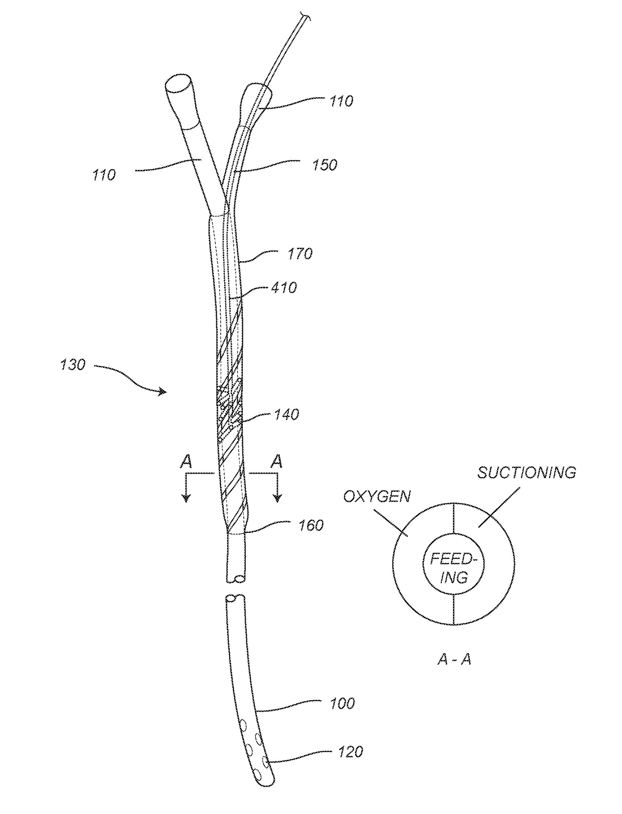
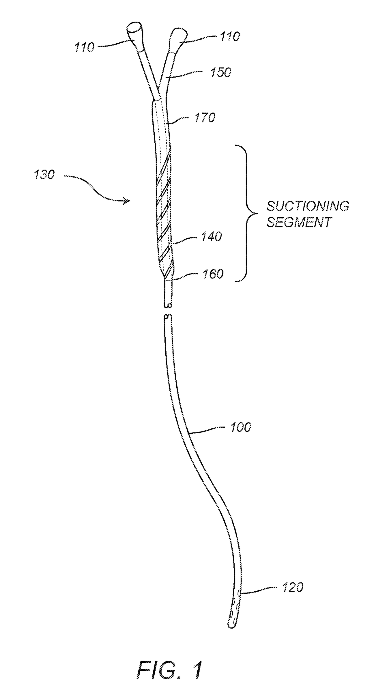
[0061] A suction tube for suctioning a patient's pharynx, the suction tube comprising: a connection port for connecting to a vacuum source and a collector; a suction section; and an enteric tube, wherein the suction section comprises a perforations and a lumen, wherein the perforations is configured to be in fluid communication with the lumen, wherein the lumen is configured to be in fluid communication with the connection port, wherein the suction section is configured to be connectable to outer portion of the enteric tube, wherein when the suction section is configured to be connectable to the enteric tube, and wherein the lumen is retained between the outer portion of the enteric tube and inside of the suction section.

[0062]Embodiment 2. The suction tube of embodiment 1, wherein the perforations comprise slots that are formed along the suction section in a longitudinally sloped orientation.

[0063]Embodiment 3. The suction tube of embodiment 1, wherein the suction section is slidab...

embodiment 9

[0069] A combination suction and enteric feeding system for feeding a patient and suctioning the patient's pharynx comprising: an enteric tube comprising an enteric lumen, an opening at a distal end of the enteric tube, a first connection port for connecting the enteric tube to a food source; and a suction tube connected to at least a portion of the enteric tube, wherein the suction tube comprises: a second connection port for connecting the suction tube to a vacuum source; and a suction lumen, wherein the suction lumen is configured to be in fluid communication with the second connection port and perforations that open to outside of the suction tube.

[0070]Embodiment 10. The system of embodiment 9, wherein the suction lumen is formed between outside of the enteric tube and inside of the suction tube in a coaxial relationship.

[0071]Embodiment 11. The system of embodiment 9, wherein the suction lumen is adjacent to and not coaxial with the enteric lumen.

[0072]Embodiment 12. The system...

embodiment 15

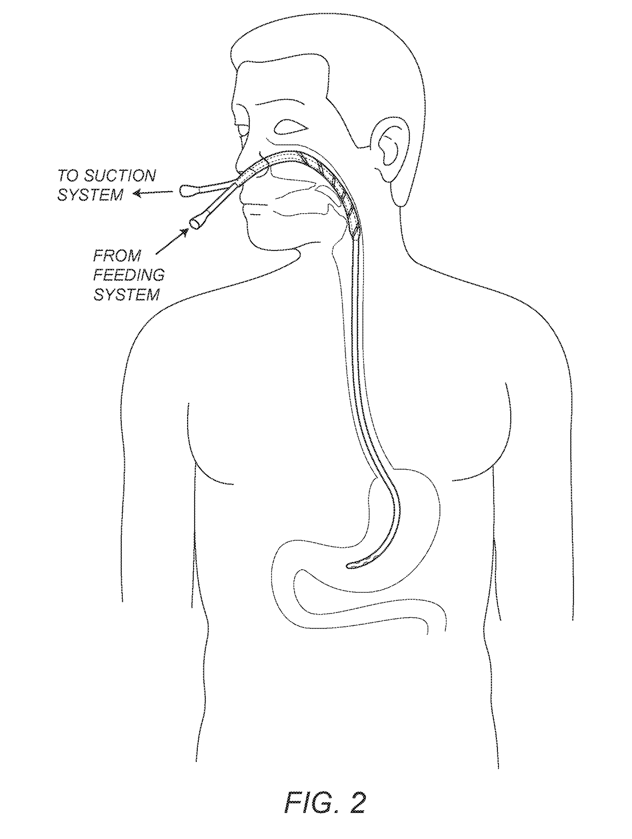
[0075] A method of suctioning a patient's pharynx while feeding the patient comprising: connecting a suction tube to an enteric tube to form a lumen around outer portion of the enteric tube and to form an airtight seal around the enteric tube, wherein the suction tube comprises: perforations that are in fluid communication with the lumen and a connector port that is configured to be in fluid communication with the lumen; inserting the connected suction and enteric tubes into the patient so that a distal end of the enteric tube is positioned in the patient's stomach and the perforations are positioned inside the patient's pharynx; connecting the suction tube to a vacuum source; and initiating suction of the vacuum source to aspire the patient's pharynx and remove secretions in the patient's pharynx.

[0076]Embodiment 16. The method of embodiment 15, wherein the enteric tube is configured to be removably connected to a food source so that food can be inserted directly into the patient's...


PUM


Abstract

The disclosed device, method, and system provide a simple, safe, effective and widely applicable method-device to suction the throat in patients with absence or incompetent swallowing reflexes. For instance, disclosed is a combination enteric feeding and pharyngeal suctioning tube that both pumps food to a patient's stomach while suctioning a patient's throat to remove pharyngeal secretions. The suctioning tube may be an attachment to a feeding tube that terminates just at or above the esophageal opening when connected to the feeding tube. The end of the pharyngeal suctioning tube may include perforations that allow the pharyngeal secretions to be vacuumed into the suctioning portion for removal.

Description

FIELD OF THE DISCLOSURE[0001]The present disclosure is directed to a device, a method, and a system for suctioning of the pharynx and enteric access for feeding and suctioning of the gastrointestinal tract and supply of oxygen to the upper airways and lungs. More particularly, the present disclosure relates to a device, a method, and a system for pumping food to a patient's stomach while suctioning a patient's throat to remove pharyngeal secretions.BACKGROUND OF THE DISCLOSURE[0002]The following description includes information that may be useful in understanding the present disclosure. It is not an admission that any of the information provided herein is prior art or relevant to the presently claimed disclosure, or that any publication specifically or implicitly referenced is prior art.[0003]Patients with a wide variety of illnesses (i.e., after a brain bleed, surgery, cancer, and the like) as well as individuals with chronic disorders of speech or swallowing motoric (i.e., after s...

Claims


Application Information

Patent Timeline
Patent Type & Authority Applications(United States)
IPC IPC(8): A61M1/00A61J15/00A61M16/10
CPCA61M1/0084A61M1/0086A61J15/0073A61M1/0023A61M16/1005A61J15/003A61M2210/1046A61M2207/00A61M2210/1028A61M2202/0208A61M2210/1053A61M2205/3303A61M1/00A61J15/0003A61M2210/005A61M2210/0625A61M2210/065A61J15/0096A61M1/85A61M1/84
Inventor ROSENGART, AXEL
Owner CEDARS SINAI MEDICAL CENT
Learn More