

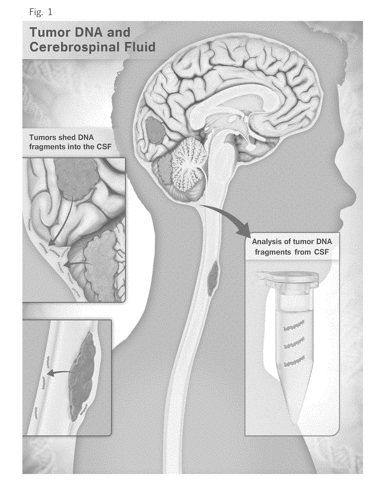
Detection of tumor-derived DNA in cerebrospinal fluid
a cerebrospinal fluid and tumor-derived technology, applied in the field of nucleic acid assays, can solve the problems of slow change, non-specific anatomic changes detected by imaging modalities, and difficult to distinguish between treatment effect and cancer growth
- Summary
- Abstract
- Description
- Claims
- Application Information
AI Technical Summary
Benefits of technology
Problems solved by technology
Method used
Image

Examples
example 1
Materials and Methods
[0033]Patient Samples. All samples were collected after approval was obtained from the
[0034]Johns Hopkins Institutional Review Board and informed consent was provided. Whole blood and cerebrospinal fluid were collected at the time of surgery, prior to surgical manipulation of the tumor. A white blood cell (WBC) pellet was prepared from the blood sample after hypotonic lysis of red blood cells via centrifugation at 200 g. CSF was frozen in its entirety at −80° C. until DNA purification, and the entire volume of CSF (cells plus fluid) was used for DNA purification. The amount of CSF used averaged 4.8 ml (range 0.75 to 10 mL). When fresh tumor tissue from surgical specimen was available, it was immediately frozen at −80° C. When frozen tissue was not available, formalin-fixed, paraffin-embedded (FFPE) tissues were used for DNA purification. In either case (fresh-frozen or FFPE) tumors were macro-dissected to ensure neoplastic cellularity exceeding 50%. DNA was puri...
example 2
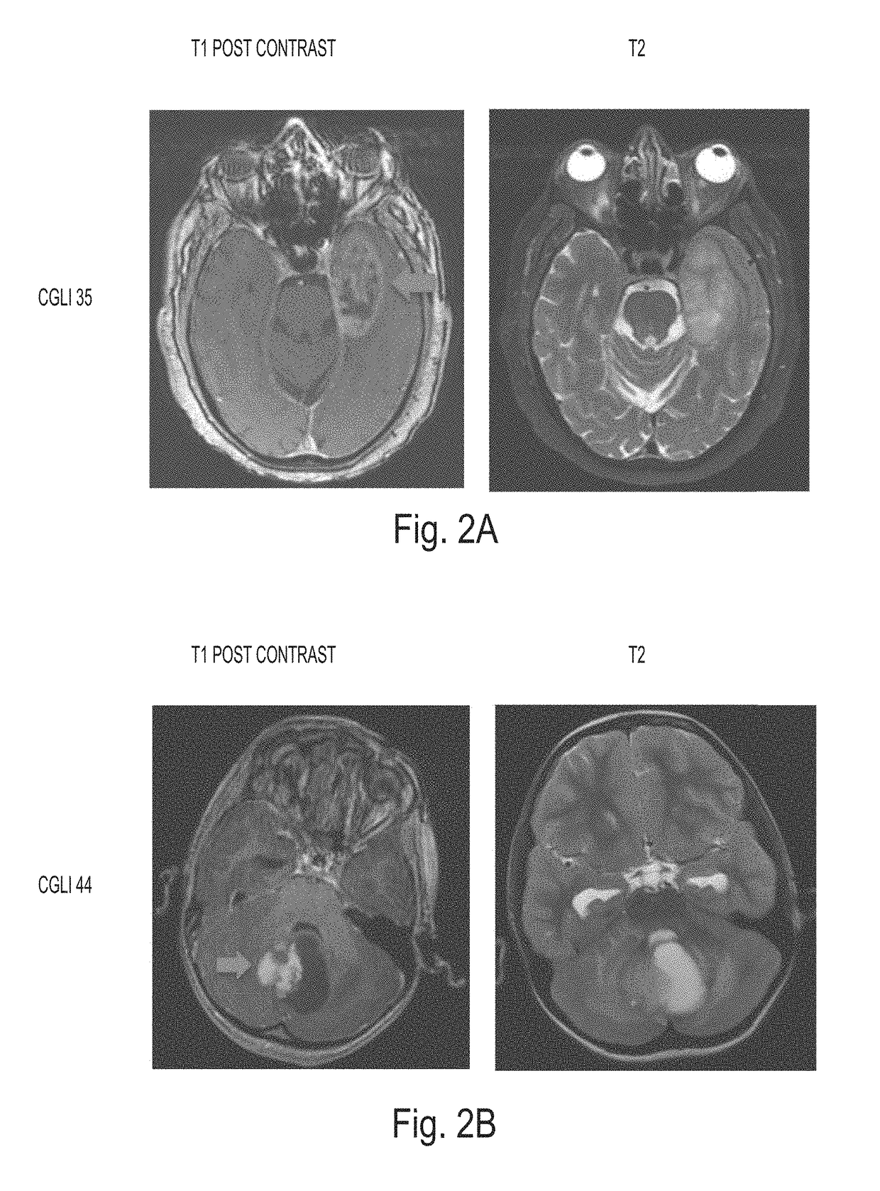
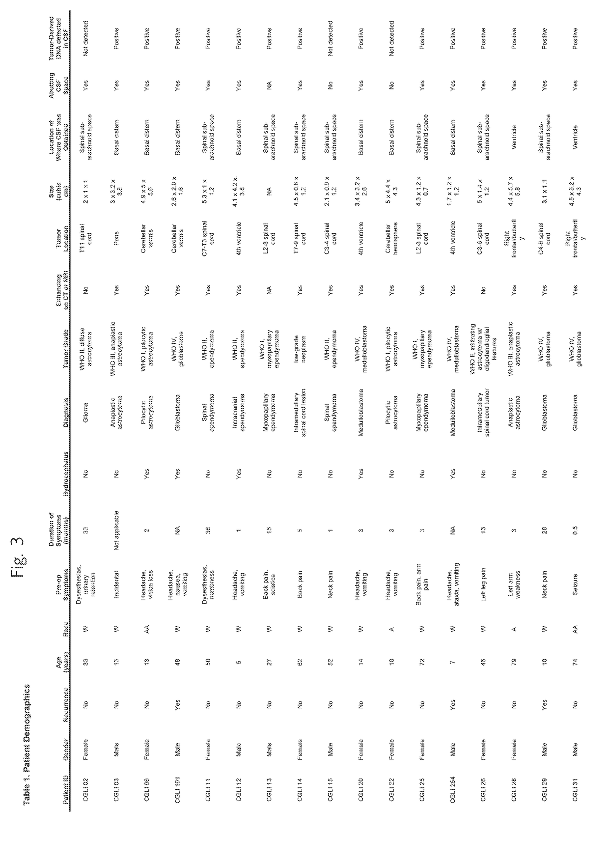
[0038]Patient and tumor characteristics. Thirty-five patients with CNS cancers were enrolled in this study. Their age, gender, race, and pre-operative symptoms are listed in Table 1, together with their tumor characteristics. Six patients had medulloblastomas and twenty-nine had gliomas. Seven, nine, two, and seventeen of the tumors were classified as WHO Grades I, II, III, and IV, respectively. Twenty-nine (83%) of the 35 patients provided CSF during the initial surgery, while the remaining six (17%) did so during a repeat resection. The tumors were distributed throughout the brain and spinal cord, with fourteen arising in the posterior fossa (including six medulloblastomas), eight in the supratentorial compartment of the brain, and thirteen in the spinal cord (Table 1).
example 3
[0039]Identification of somatic mutations. At least one mutation was identified in each of the 35 tumors analyzed using a tiered approach (targeted sequencing followed by whole-exome sequencing) described in the Materials and Methods section.
[0040]With the targeted sequencing approach, we identified mutations in thirteen tumors. The mutations in these samples occurred in TP53 (n=5), IDH1 (n=2), and the TERT promoter (n=6) (Table 2). In the remaining 22 tumors, whole-exome sequencing was used to identifiy at least one mutation per sample. Genes mutated in these samples included well-known drivers, such as NF2, PIK3R1, PTCH1, and PTEN (18). The fractions of mutant alleles in tumors were generally high, averaging 46% (with standard deviation (SD) of 18%). This is consistent with the expected early development of driver gene mutations during tumor evolution and the presence of non-neoplastic cells in all tumors, even macrodissected ones such as the samples used here. All mutations ident...
PUM
| Property | Measurement | Unit |
|---|---|---|
| Fraction | aaaaa | aaaaa |
| Fraction | aaaaa | aaaaa |
| Fraction | aaaaa | aaaaa |
Abstract
Description
Claims
Application Information