Noninvasive, label-free, in vivo flow cytometry using speckle correlation technique

a correlation technique and flow cytometry technology, applied in the field of flow cytometry, can solve the problems of limiting the depth at which we can reliably perform flow cytometry, reducing the accuracy of flow cytometry, and challenging optical imaging

Active Publication Date: 2019-01-03
CALIFORNIA INST OF TECH
View PDF0 Cites 9 Cited by
  • Summary
  • Abstract
  • Description
  • Claims
  • Application Information

AI Technical Summary

Benefits of technology

[0030]14. The apparatus of one or any combination of the embodiments 1-13, wherein the sample holder includes a clamp clamping the blood vessel so as to control a velocity of blood flow through the blood vessel.

Problems solved by technology

Optical imaging is challenging in turbid media, where multiple scattering of light causes a degradation of resolution and limits the depths at which we can reliably image (<1 mm in biological tissue) without having to resort to destructive optical clearing or sectioning techniques [1].
An issue with methods that rely on detecting only the minimally scattered photons is the maximum achievable depth of penetration, since the chance of detecting a quasi-ballistic photons decreases exponentially with increasing depth.
However, SCI methods are limited to working in dark-field scenarios, with sparsely-tagged objects [14], since the detected light must consist solely of light arising from the object.

Method used


Image

  • Noninvasive, label-free, in vivo flow cytometry using speckle correlation technique
  • Noninvasive, label-free, in vivo flow cytometry using speckle correlation technique
  • Noninvasive, label-free, in vivo flow cytometry using speckle correlation technique

Examples


first example

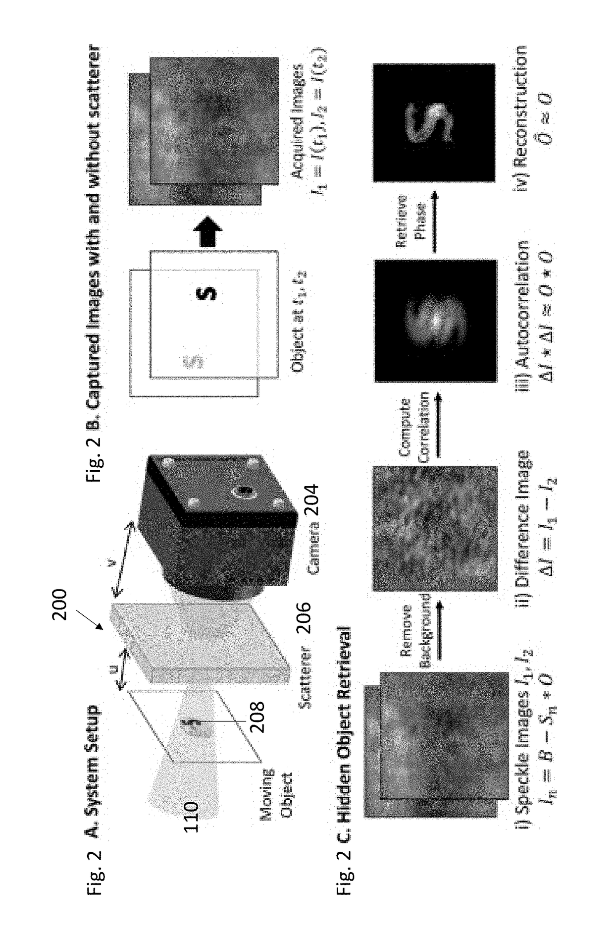
[0052]FIG. 2A illustrates an example apparatus 200 for performing SCFC, illustrating the scattered light 202 is detected by a high-resolution camera 204 that is placed at a distance v from the scattering media 206 and wherein the moving object 208 is at a distance u from the scatterer. The resultant object will have an image size dictated by the magnification of the system, M=−v / u.

[0053]FIG. 2B illustrates captured images with and without the scatterer.

[0054]FIG. 2C illustrates the hidden object can be retrieved from the seemingly random speckle images by taking advantage of inherent angular correlations in the scattering pattern. FIG. 2C(i) shows each captured image In consists of a background, B, subtracted by the imaged object, where the imaged object is the convolution of the PSF of the scattering media, S, and the object pattern, O. FIG. 2C(ii) shows that, although the background signal dominates over the object, it can be subtracted out by taking the difference between the two...

case 1

Travels Distance Where C(Δx)Δ1

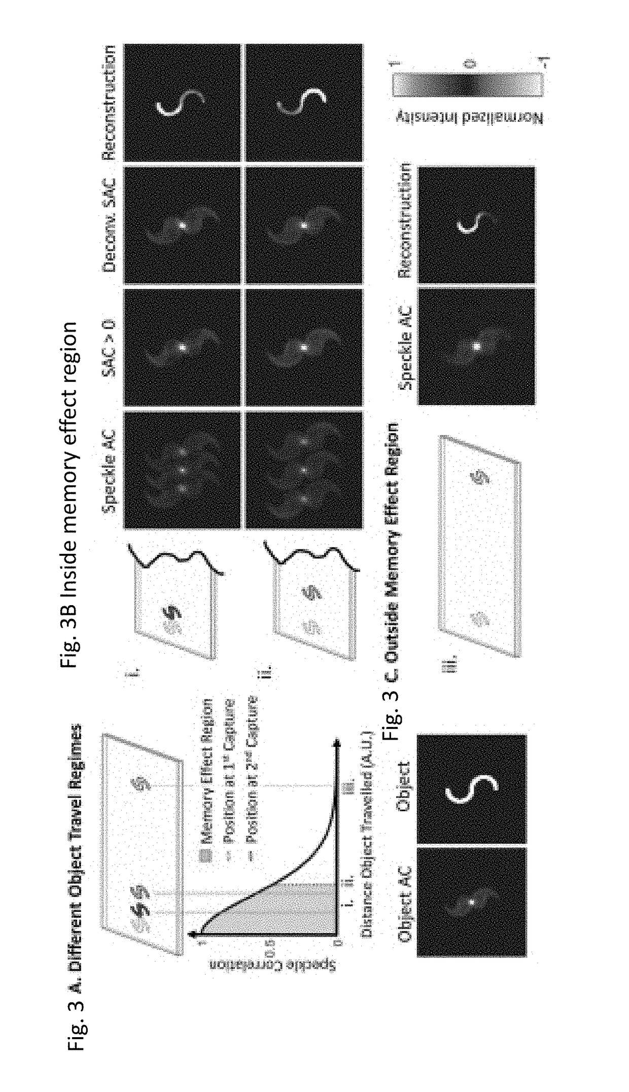
[0058]In the case where the object travels a small distance (such that C(Δx)≈1), we have

S2(xi)≈S1(xi+Δxi)  (7)

[0059]where x=(x, y), xi=(xi, yi) are coordinates in the object plane and image plane respectively, Δx is the distance the object traveled in the object plane, and Δxi=MΔx. We can equivalently consider the PSF to be the same in both captures and have the object travel between captures.

[0060]That is,

O2=O(xi+Δxi),  (8)

ΔI=S*[O(xi)−O(xi+Δxi)],  (9)

and

ΔI*ΔI=2A(xi)−A(xi+Δxi)−A(xi−Δxi).  (10)

[0061]where A=O*O is the object, autocorrelation (OAC). The SAC contains three copies of the OAC: a positive copy centered at x=(0, 0), and two negative copies shifted by an amount commensurate with the object travel distance [FIG. 5B, “Speckle AC”)].

[0062]Since C(Δx)≈1 when Δx≈0, the object may travel a distance shorter than the extent of its autocorrelation. In this case, the SAC will yield positive and negative copies of the OAC that overlap [FIG. 3Bi]. The OAC ...

case 2

Travels Distance Where C(Δx)>0.5

[0063]In the regime where the object travels within the angular ME range (C(Δx)>0.5), S1 and S2 are correlated. To highlight the impact of the degree of correlation C(Δx) on the SAC, we can mathematically represent S2 as:

S2=C(Δx)S1(xi+Δxi)+√{square root over (1−[C(Δx)]2)}S,  (11)

[0064]where S is a speckle intensity pattern that is uncorrelated with S1. The scatter PSFs in the equation above are mean-subtracted speckle intensities. Representing S2 in the form above allows us to preserve speckle intensity statistics (that is, the speckle intensity variance and mean satisfy V[S1]=V[S2] and E[S1]=E[S2] respectively.)

[0065]Using Eq. (11), Eqs. (4) and (5) become

ΔI=(S1−C(Δx)S1(x1+Δxi)−√{square root over (1−[C(Δx)]2)}S)*O  (12)

and

ΔI*ΔI≈2A(xi)−C(Δx)A(xi±Δxi)+√{square root over (1−[C(Δx)]2)}×noise,  (13)

[0066]where the last equation follows from noting that the speckle fields are a delta-correlated process and that the cross-correlation of two uncorrelated spe...


PUM

PropertyMeasurementUnit
decorrelation timeaaaaaaaaaa
depthsaaaaaaaaaa
timeaaaaaaaaaa

Abstract

A system and method for performing speckle correlation flow cytometry (SCFC). By subtracting out the stationary background when shining light through a sample (e.g., a vessel within a biological tissue), light only scattered by the desired targets (e.g., cells) can be captured and different types of targets (e.g., cells) can be distinguished by the autocorrelation of the speckle pattern. In this way, the targets (e.g., cells) can be classified and counted based on the features of their speckle correlations. The technique can be applied not only for noninvasive, label-free, in vivo CTC counting but also for counting other types of blood cells such as white blood cells or red blood cells.

Description

CROSS REFERENCE TO RELATED APPLICATIONS[0001]This application claims the benefit under 35 U.S.C. Section 119(e) of co-pending and commonly-assigned U.S. Provisional Patent Application Ser. No. 62 / 527,237, filed on Jun. 30, 2017, by Edward H. Zhou and Michelle Cua, entitled “NONINVASIVE, LABEL-FREE, IN VIVO FLOW CYTOMETRY USING SPECKLE CORRELATION TECHNIQUE,” CIT-7811), which application is incorporated by reference herein.STATEMFNT REGARDING FEDERALLY SPONSORED RESEARCH AND DEVELOPMENT[0002]This invention was made with government support under Grant No. NS090577 awarded by the National Institutes of Health. The government has certain rights in the invention.BACKGROUND OF THE INVENTION1. Field of the Invention[0003]This invention relates to a method and system for imaging and / or performing flow cytometry.2. Description of the Related Art[0004](Note: This application references a number of different publications as indicated throughout the specification by one or more reference number...

Claims


Application Information

Patent Timeline
Patent Type & Authority Applications(United States)
IPC IPC(8): G06K9/62G01N15/14A61B5/1455A61B5/145A61B90/20G06T7/00G06T7/246G06V10/141
CPCG06K9/628G01N15/1429G01N15/1404A61B5/1455A61B5/14535A61B90/20G06T7/0012G06T7/246G06T2207/30242G06T2207/30101G06T2207/30096G01N2015/1454G01N2015/1006G01N15/1459G06T2207/10016G06T2207/30024G06T2207/30104G06T7/254G06V20/69G06V10/141G06F18/2431
Inventor ZHOU, EDWARD H.CUA, MICHELLE
Owner CALIFORNIA INST OF TECH
Why Eureka
Try Eureka