Exosome-Total-Isolation-Chip (ExoTIC) Device for Isolation of Exosome-Based Biomarkers

a biomarker and exosome technology, applied in the field of identification of extracellular vesicle or exosome-based biomarkers, can solve the problems of large sample volume, difficult to translate exosomes as cancer biomarkers into clinical practice, and large sample volume, etc., and achieve the effect of cost-effectiveness

Active Publication Date: 2019-02-14
THE BOARD OF TRUSTEES OF THE LELAND STANFORD JUNIOR UNIV
View PDF0 Cites 2 Cited by
  • Summary
  • Abstract
  • Description
  • Claims
  • Application Information

AI Technical Summary

Benefits of technology

[0027]It is contemplated that there are many potential advantages of the ExoTIC device over existing methods, devices, and / or materials. In comparison to traditional means, the ExoTIC device is both time and labor saving and very cost effective. Still yet, as will be discussed below, it yields greater amount of exosomes from a sample that other methods. It is usable with a wide range of sample volumes, including from 10 μL to 50 mL and is suitable for use on all types of biological samples (including, but not limited to, plasma, serum, urine, saliva, culture media, and ascitic fluid). The device can process in series and parallel configurations larger volumes than 50 mL. Still further, it is contemplated that it may be integrated with microfluidic chip devices. Moreover, because of the manner in which it operates the ExoTIC can provide size-specific exosome isolation.

Problems solved by technology

To date, translating exosomes as cancer biomarkers into clinical practice has proven challenging as a reliable source for diagnosis.
Existing exosome purification technologies (ultracentrifugation, multi-step filtration, antibody conjugated magnetic beads or polyethylene glycol based precipitation, see the left three panels on the bottom of FIG. 1) are often expensive, time-consuming, require large sample volumes and frequent manual handling, and / or result in poor yields and unpredictable purity of exosome preparations, especially when derived from clinical plasma samples.
This inability to efficiently purify and isolate exosomes has proven to be a major impediment to the application of exosomes for drug delivery and diagnostics.

Method used


Image

  • Exosome-Total-Isolation-Chip (ExoTIC) Device for Isolation of Exosome-Based Biomarkers
  • Exosome-Total-Isolation-Chip (ExoTIC) Device for Isolation of Exosome-Based Biomarkers
  • Exosome-Total-Isolation-Chip (ExoTIC) Device for Isolation of Exosome-Based Biomarkers

Examples


Embodiment Construction

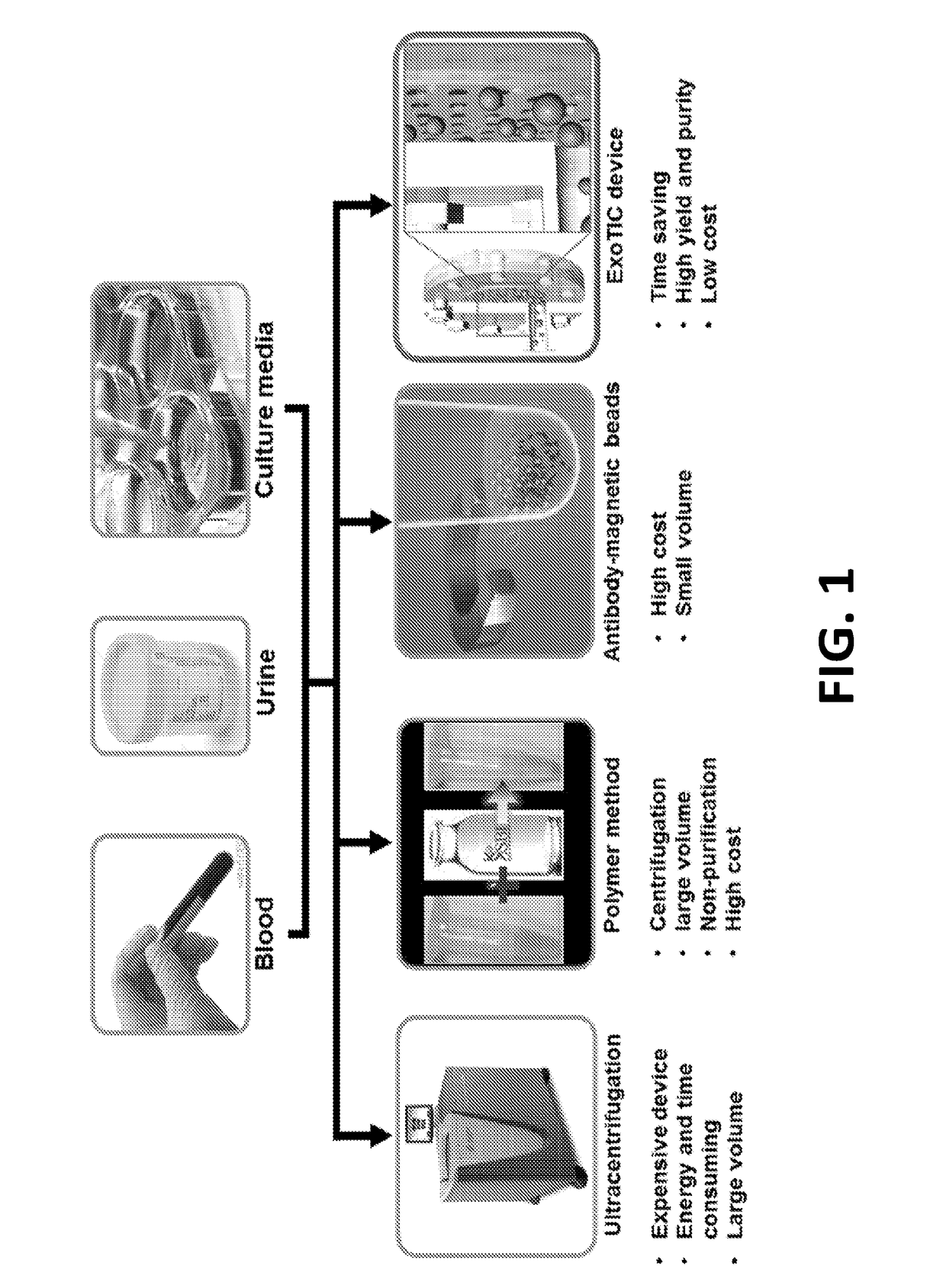
[0045]As noted above, exosomes carry biological analytes (for example, proteins, metabolites, and nucleotides) that reflect their cell type of origin. Due to their important role as messengers in cell-cell communication, exosomes are emerging both as candidate biomarkers in liquid biopsies and as targeted drug delivery vehicles for therapeutics. However, a major challenge hindering the routine clinical use of exosomes is the lack of simple and inexpensive, yet robust and reproducible methods for their isolation and enrichment. Table 1 below, provides a brief summary of each of the existing exosome purification technologies and compares them with the ExoTIC device disclosed herein.

TABLE 1Comparison of different methods for exosome isolationExoExoMethodsSample typesVolumeYieldPurityTimeUltracen-Culture media>30mLLowHighDaystrifugationAscitic fluid(UC)UrineFilterCulture media60mLLowLowHoursmethodPolyethyleneCulture media1-5mLLowLowHoursglycol (PEG)Plasma / serum60-300μLAscitic fluid1-5mL...


PUM

PropertyMeasurementUnit
pore diameteraaaaaaaaaa

Abstract

A device (“the ExoTIC device”) for the isolation of extracellular vesicles from an extracellular vesicle-containing sample in which the sample is flowed through a membrane in a flow chamber to capture and purify the extracellular vesicles on the membrane. The extracellular vesicles may be washed and collected and utilized in any one of a number of ways including, but not limited to, identifying biomarkers of a disease, identifying the presence of a biomarker in a patient to determine whether a patient has a disease, and therapeutically treating existing diseases by re-introducing the extracellular vesicles, potentially modified, back into a body.

Description

CROSS-REFERENCE TO RELATED APPLICATIONS[0001]This application claims the benefit of U.S. Provisional Patent Application No. 62 / 289,795 entitled “Exosome-Total-Isolation-Chip (ExoTIC) Device for Identification of Exosome-Based Biomarkers” filed Feb. 1, 2016, the contents of which are incorporated by reference herein in its entirety for all purposes.STATEMENT OF FEDERALLY SPONSORED RESEARCH OR DEVELOPMENT[0002]This invention was made with Government support under contract CA199075 and DE024971 awarded by the National Institutes of Health. The Government has certain rights in the invention.BACKGROUND[0003]This disclosure relates to the identification of extracellular vesicle or exosome-based biomarkers and, in particular, to devices and methods of using these devices for isolation of extracellular vesicle or exosome-based biomarkers.[0004]Exosomes, which are small (generally, 30-180 nm) cell-derived vesicles, are abundantly secreted by different types of cells into the extracellular fl...

Claims


Application Information

Patent Timeline
Patent Type & Authority Applications(United States)
IPC IPC(8): G01N33/50B01L3/00G01N33/53
CPCG01N33/5091B01L3/502715G01N33/5308B01L3/50273B01L3/502761G01N2800/52G01N2800/56G01N33/50G01N33/5005
Inventor LIU, FEIDEMIRCI, UTKANGAMBHIR, SANJIV SAMNAIR, VISWAM S.
Owner THE BOARD OF TRUSTEES OF THE LELAND STANFORD JUNIOR UNIV
Why Eureka
Try Eureka