
Contrast Dose Reduction for Medical Imaging Using Deep Learning

Active Publication Date: 2019-04-11
THE BOARD OF TRUSTEES OF THE LELAND STANFORD JUNIOR UNIV
View PDF0 Cites 30 Cited by
  • Summary
  • Abstract
  • Description
  • Claims
  • Application Information

AI Technical Summary

Benefits of technology

The present invention offers techniques to improve the image quality of medical imaging without using a higher dose of contrast. These techniques can be applied to various imaging modalities such as MRI, angiography, computed tomography, and ultrasound. The invention can predict a full-dose contrast agent image from a low-dose contrast agent image and a pre-dose image with high diagnostic quality. These techniques yield significant improvements over the conventional low-dose images with over 5 dB PSNR gains, 11% SSIM increased, and improvements in ratings on image quality and contrast enhancement.

Problems solved by technology

Significantly, naively amplifying the contrast enhancement of a 1 / 10 low-dose CE-MRI by a factor of ten results in poor image quality with widespread noise and ambiguous structures.

Method used


Image

  • Contrast Dose Reduction for Medical Imaging Using Deep Learning
  • Contrast Dose Reduction for Medical Imaging Using Deep Learning
  • Contrast Dose Reduction for Medical Imaging Using Deep Learning

Examples


Embodiment Construction

[0022]Embodiments of the present invention provide a deep learning based diagnostic imaging technique to significantly reduce contrast agent dose levels while maintaining diagnostic quality for clinical images.

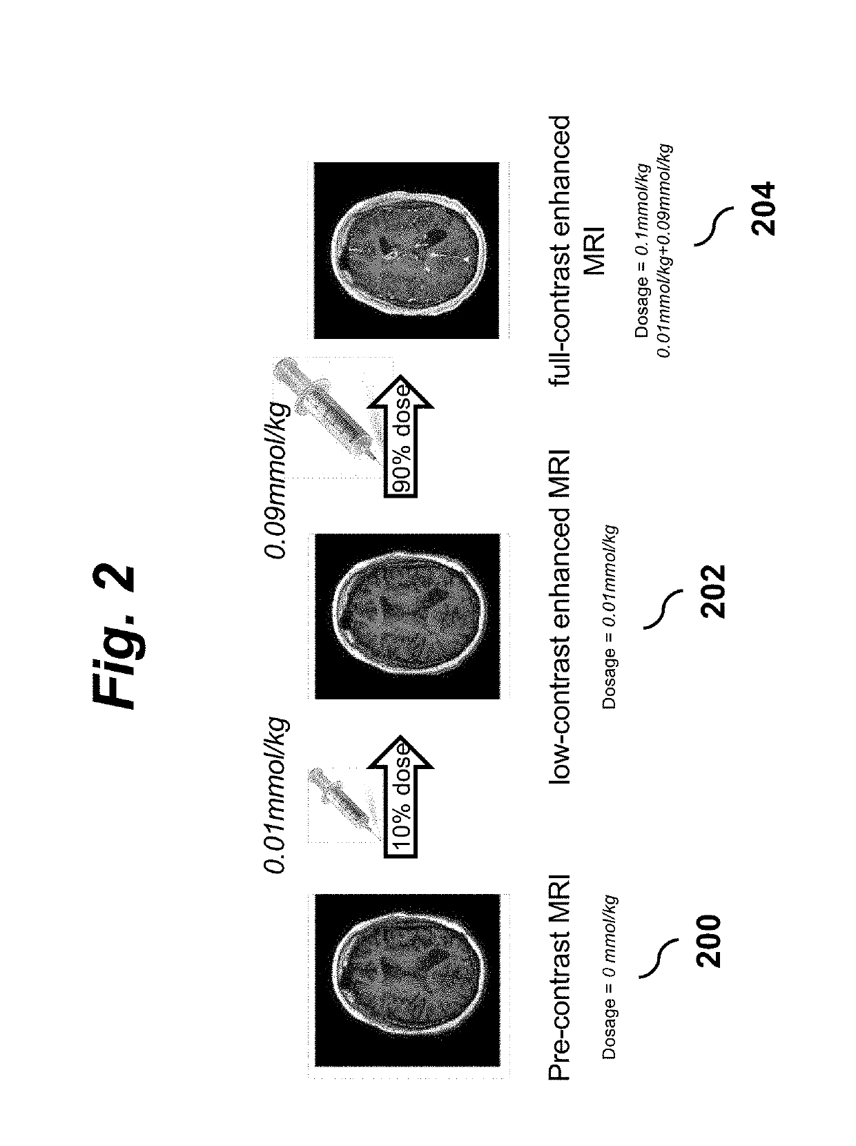
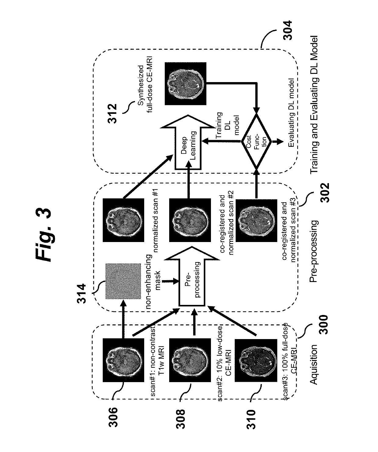
[0023]Detailed illustrations of the protocol and procedure of an embodiment of the invention are shown inFIG. 1 and FIG. 2. Although the embodiments described below focus on MRI imaging for purposes of illustration, the principles and techniques of the invention described herein are not limited to MRI but are generally applicable to various imaging modalities that make use of contrast agents.

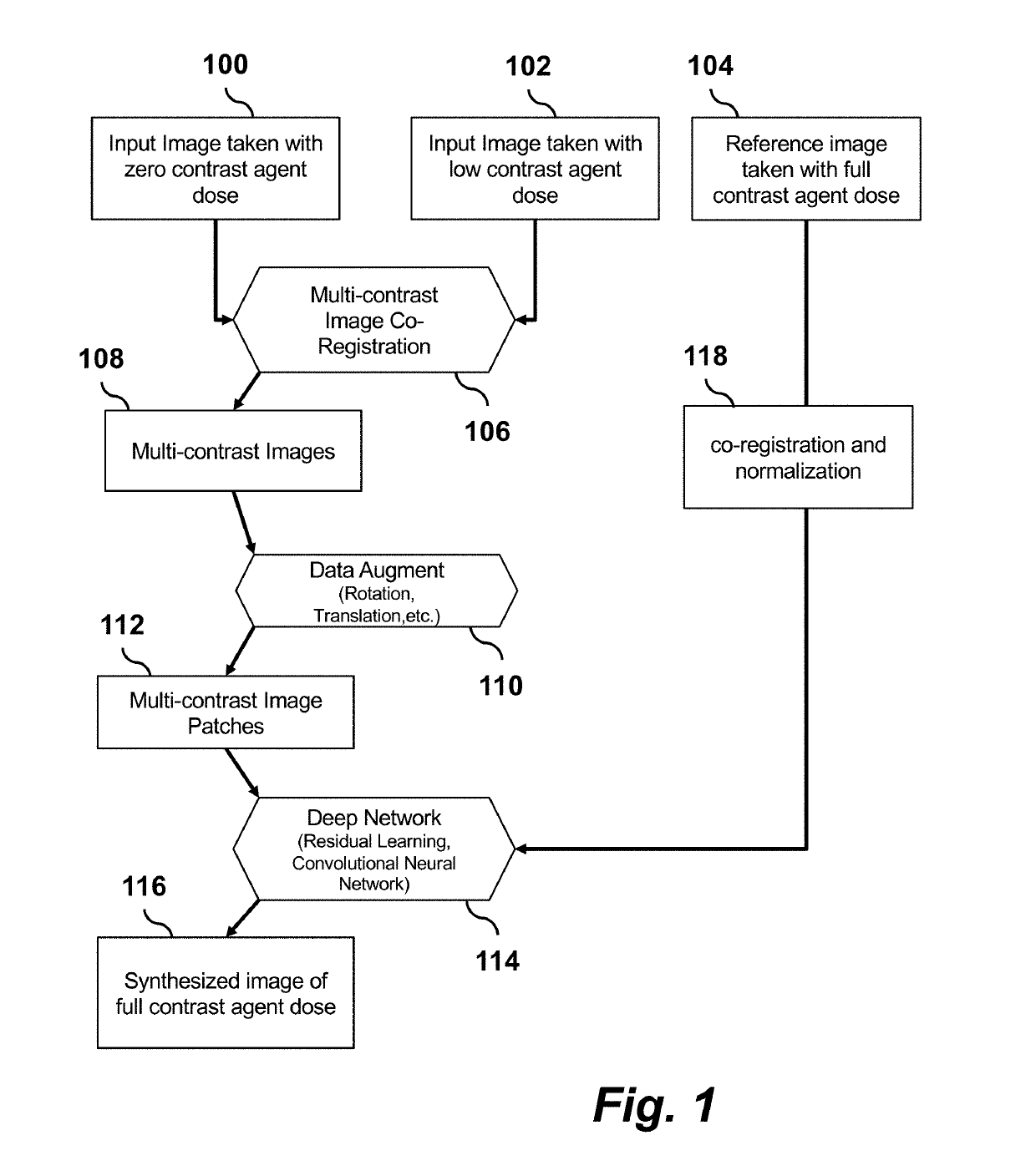
[0024]FIG. 1 is a flow chart showing a processing pipeline for an embodiment of the invention. A deep learning network is trained using multi-contrast images 100, 102, 104 acquired from scans of a multitude of subjects with a wide range of clinical indications. The images are pre-processed to perform image co-registration 106, to produce multi-contrast images 108, and data augmentation 110...


PUM


Abstract

A method for diagnostic imaging with reduced contrast agent dose uses a deep learning network (DLN) [114] that has been trained using zero-contrast [100] and low-contrastimages as input to the DLN and full-contrast images [104] as reference ground truth images. Prior to training, the images are pre-processed [106, 110, 118] to co-register and normalize them. The trained DLN [114] is then used to predict a synthesized full-dose contrast agent image [116] from acquired zero-dose and low-dose images.

Description

CROSS-REFERENCE TO RELATED APPLICATIONS[0001]This application claims priority from U.S. Provisional Patent Application 62 / 570,068 filed Oct. 9, 2017, which is incorporated herein by reference.STATEMENT REGARDING FEDERALLY SPONSORED RESEARCH OR DEVELOPMENT[0002]None.FIELD OF THE INVENTION[0003]This invention relates generally to medical diagnostic imaging. More specifically, it relates to imaging techniques that use contrast agents.BACKGROUND OF THE INVENTION[0004]Many types of medical imaging use contrast agents to enhance the visualization of normal and abnormal structures. Examples include conventional angiography, fluoroscopy, computed tomography (CT), ultrasound, and magnetic resonance imaging (MRI). It is often desirable to reduce the contrast agent dose in order to increase the safety of these agents. However, reduced dose reduces desired imaging enhancements, so this has not been possible.[0005]For example, MRI is a powerful imaging technique providing unique information to d...

Claims


Application Information

Patent Timeline
IPC IPC(8): G06T7/00G06T3/60G06N3/08
CPCG06T7/0012G06T3/60G06N3/08G06T2207/10081G06T2207/10088G06T5/002G16H30/40G06T2207/10121G06T2207/20081G06T2207/10072G06T2207/10132G06T2207/20084G06T2207/30004G16H50/20G06N3/084G06T5/50G06T2207/30016G06T2207/10116G06N3/048G06N3/045G06T5/60G06T7/50G06T2207/20221G06T5/70
Inventor ZAHARCHUK, GREGGONG, ENHAOPAULY, JOHN M.
Owner THE BOARD OF TRUSTEES OF THE LELAND STANFORD JUNIOR UNIV
Learn More