Scaffold-free 3D bioprinting of porcine cells
a 3d bioprinting and porcine cell technology, applied in the field of scaffold-free 3d bioprinting of porcine cells, can solve the problems of strong immunological response and survival-limiting thrombocytopenia of xenotransplant recipients, and it is unknown what genetic combination will be best to reduce immunological and coagulation responses
- Summary
- Abstract
- Description
- Claims
- Application Information
AI Technical Summary
Benefits of technology
Problems solved by technology
Method used
Image
Examples
example 1
Generating a Scaffold-Free 3D Bio-Assembled Genetically Engineered Pig Tissue / Liver
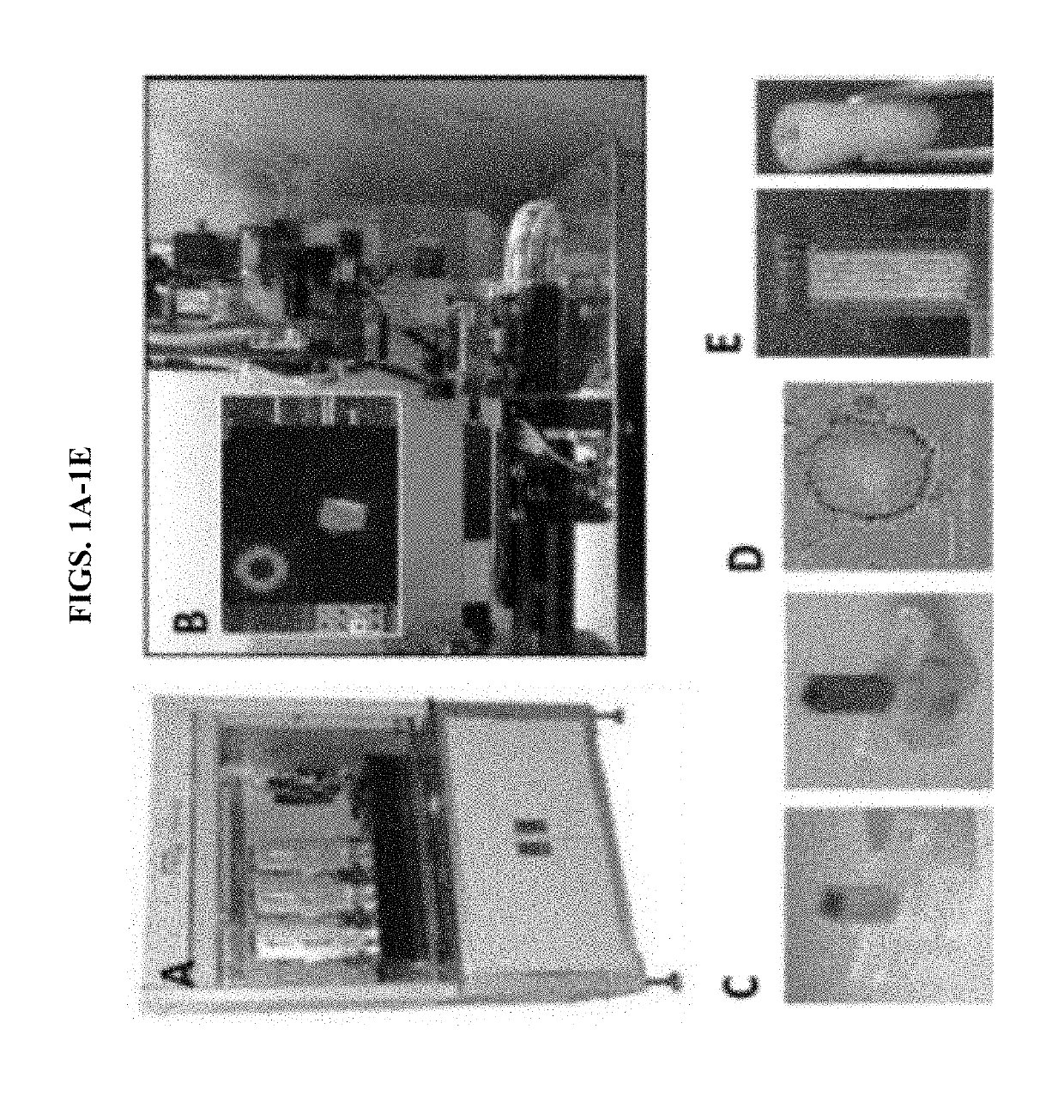
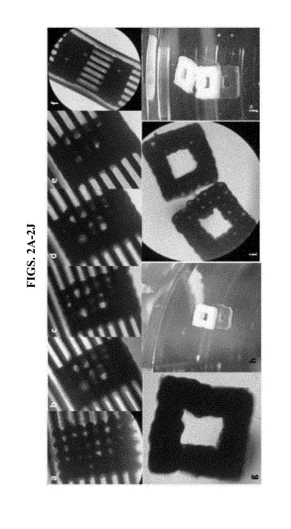
[0066]Recently, a high-throughput instrument that is capable of assembling spheroids with precision has been developed: a Regenova bioprinter by Cyfuse Biomedical (Japan) (FIG. 1). In general, robotic platforms can assemble cell spheroids into predesigned contiguous structures with submillimeter-level three-dimensional (3D) precision, using an arrangement of microneedles as support. Using a Regenova bioprinter, we printed scaffold-free 3D constructs using wild-type (WT) and genetically-engineered pig cells onto Kenzan microneedles. Referring to FIGS. 2A-2J, we bioprinted spheroids comprising wild-type porcine fibroblasts and liver derived cells (LDCs). The bioprinted spheroids fused to form a three-dimensional construct of cellular aggregates upon the microneedles within the first five days following printing. On day 5, we removed the 3D constructs from the microneedles. Micro-holes were visible in th...
example 2
Scaffold-Free 3D-Bioprinting of a Liver Model
[0071]This example demonstrates production and printing of hepatocyte and hepatic stellate cell (HSC) spheroids to create a SF3DBP liver model. Briefly, freshly thawed primary pig hepatocytes and immortalized pig HSC were used to generate spheroids with (i) hepatocytes alone, (ii) HSC alone, or (iii) a combination of hepatocytes and HSC. Spheroids were formed using low adhesion plates, then characterized for distance from well center, diameter, roundness, and smoothness. A column of spheroids was printed using a Regenova 3D-bioprinter. Remaining loose spheroids were incubated over two weeks for functionality assays (albumin secretion, mRNA transcription, urea clearance).
[0072]Materials and Methods
[0073]Cell culture: Hepatocytes & HSCs were isolated from the liver of a 4-month old, healthy male pig (FIG. 11). HSC were immortalized using SV40T lentivirus. Immortalized HSCs were successfully cultured.
[0074]Spheroid formation: Previously isol...
example 3
Scaffold-Free 3D-Bioprinting of Lung
[0086]Spheroids were formed using three types of porcine pulmonary cells: pulmonary vascular endothelial cells (CD31+ve), pulmonary fibroblasts, and pulmonary pneumocytes Type II. Different ratios of pulmonary vascular endothelial cells, pulmonary fibroblasts, and pulmonary pneumocytes Type II were tested to form spheroids. The most suitable spheroids were formed using the ratio 1:1:1 / 2 of pulmonary vascular endothelial cells, pulmonary fibroblasts, and pulmonary pneumocytes Type II, respectively. Total cell numbers per spheroid was about 40,000 cells. Spheroids were bioprinted 2-3 days after they were matured in 96-well U bottom plate. A special hollow model computer design was chosen, as shown in FIG. 17 and spheroids were bioprinted on temporary microneedles (FIG. 17). Starting from day 1 post-bioprinting, spheroids starting to fuse making their own extracellular matrix (FIG. 18). By day 5 post-bioprinting, a solid, fused 3D lung construct was ...
PUM
 Abstract
Description
Claims
Application Information
 - R&D Engineer
- R&D Manager
- IP Professional
- Industry Leading Data Capabilities
- Powerful AI technology
- Patent DNA Extraction
Browse by: Latest US Patents, China's latest patents, Technical Efficacy Thesaurus, Application Domain, Technology Topic, Popular Technical Reports.
© 2024 PatSnap. All rights reserved.Legal|Privacy policy|Modern Slavery Act Transparency Statement|Sitemap|About US| Contact US: help@patsnap.com


