
System and method for assessment of retinal and choroidal blood flow noninvasively using color amplification

Active Publication Date: 2019-05-30
UNIV OF KENTUCKY RES FOUND
View PDF0 Cites 8 Cited by
  • Summary
  • Abstract
  • Description
  • Claims
  • Application Information

AI Technical Summary

Benefits of technology

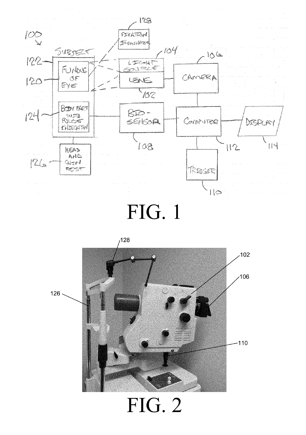
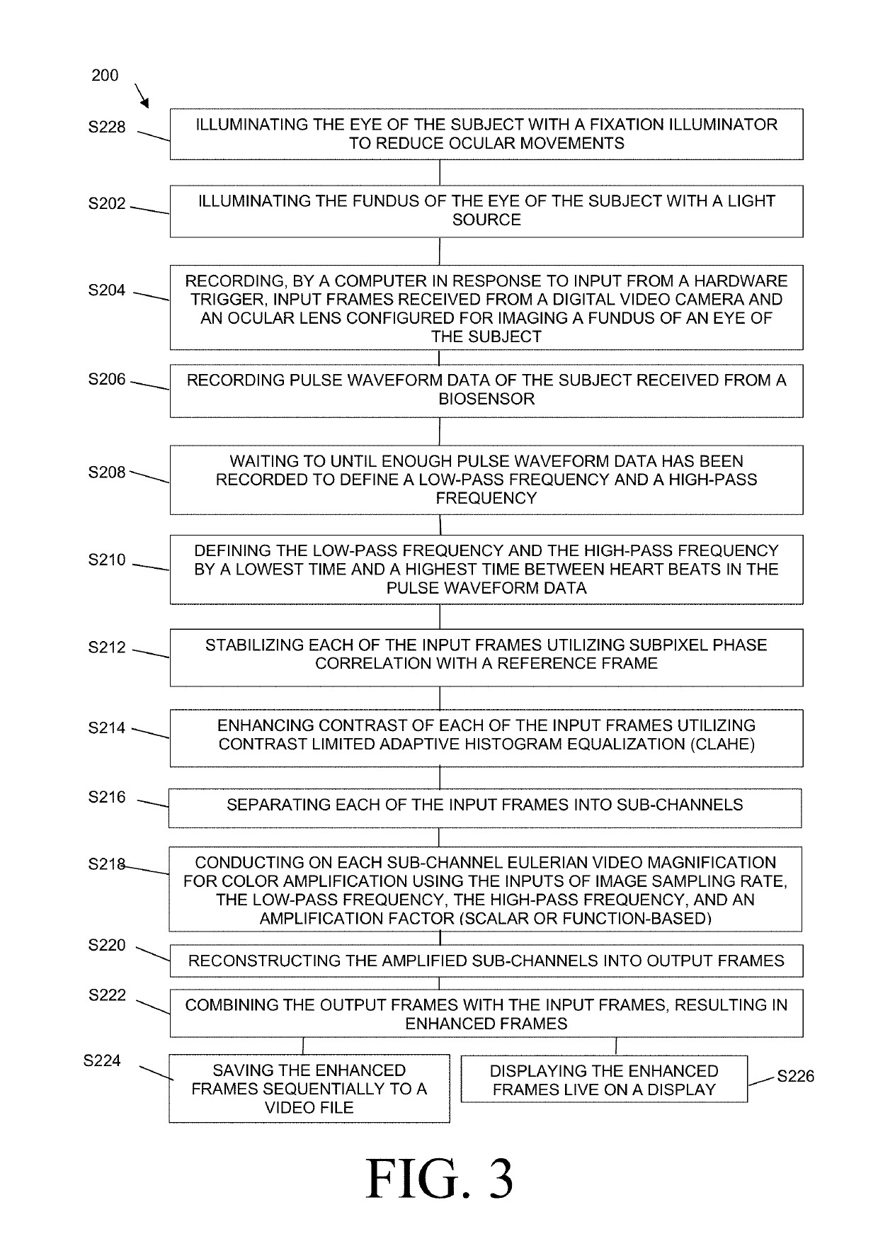
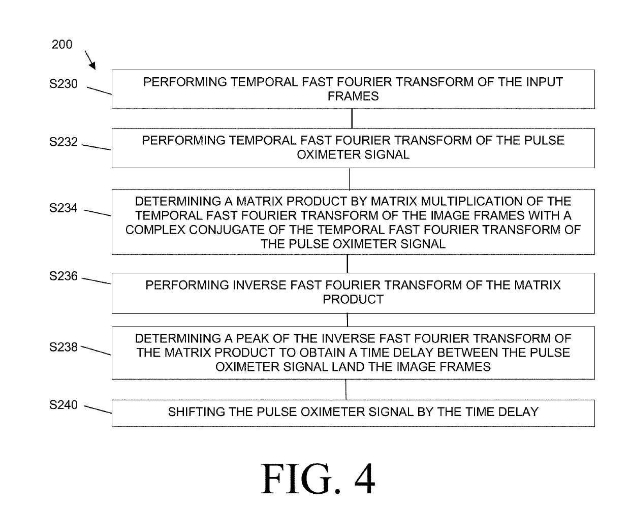
This patent describes a system and method for measuring retinal and choroidal blood flow in a subject using a digital video camera and an ocular lens. The system also includes a fixation illuminator to reduce ocular movements. The method involves recording input frames and pulse waveform data from the subject, identifying low-pass and high-pass frequencies, stabilizing each frame, enhancing contrast, separating frames into sub-channels, and conducting eulerian video magnification for color amplification. The resulting output frames are then combined with the input frames to create enhanced frames demonstrating tissue perfusion. The technical effects of this patent are improved accuracy and precision in measuring retinal and choroidal blood flow, which can be useful in the diagnosis and treatment of retinal diseases such as glaucoma.

Problems solved by technology

Despite growing evidence of choroidal abnormalities present in diabetes, it remains unclear how these changes clinically impact diabetic patients.
Although the retina itself is readily available for imaging, the RPE obscures the choroid, making it difficult to visualize using standard ophthalmic imaging techniques.
This difficulty hinders efforts in using choroidal abnormalities as a predictive factor of disease evolution and response.
Although this technique can detect gross vascular defects, it does not provide much information concerning anatomic or structural features of the choroid, and it requires static images taken sequentially over minutes.
Additionally, the ICG contrast dye is known to cause allergic reactions and requires venipuncture making the technique invasive to the patient.

Method used


Image

  • System and method for assessment of retinal and choroidal blood flow noninvasively using color amplification
  • System and method for assessment of retinal and choroidal blood flow noninvasively using color amplification
  • System and method for assessment of retinal and choroidal blood flow noninvasively using color amplification

Examples


Embodiment Construction

[0027]The details of one or more embodiments of the presently-disclosed invention are set forth in this document. Modifications to embodiments described herein, and other embodiments, will be evident to those of ordinary skill in the art after a study of the information provided herein. The information provided herein, and particularly the specific details of the described exemplary embodiments, is provided primarily for clearness of understanding and no unnecessary limitations are to be understood therefrom. In case of conflict, the specification of this document, including definitions, will control.

[0028]While the terms used herein are believed to be well understood by one of ordinary skill in the art, definitions are set forth herein to facilitate explanation of the presently-disclosed subject matter.

[0029]Unless defined otherwise, all technical and scientific terms used herein have the same meaning as commonly understood by one of ordinary skill in the art to which the presently...


PUM


Abstract

A system and method for assessing blood flow include: an ocular lens; a light source; a digital video camera; a biosensor; a trigger; and a computer. The ocular lens is for viewing a fundus of an eye. The light source is for illuminating the fundus. The digital video camera is for imaging the fundus. The biosensor is for sensing a pulse waveform. The computer is configured for: recording input frames and pulse waveform data in response to an input from the trigger; defining a low-pass frequency and a high-pass frequency from the pulse waveform data; stabilizing the input frames; enhancing contrast of the input frames; separating the input frames into sub-channels; conducting eulerian video magnification for color amplification using the inputs of image sampling rate, the low-pass frequency, the high-pass frequency, and an amplification factor; reconstructing the sub-channels into output frames; and combining the output frames with the input frames.

Description

RELATED APPLICATIONS[0001]This application claims priority to U.S. Provisional Patent Application No. 62 / 593,045, filed Nov. 30, 2017, the entire disclosure of which is incorporated herein by reference.STATEMENT REGARDING FEDERALLY SPONSORED RESEARCH OR DEVELOPMENT[0002]The presently disclosed subject matter was made with support from the U.S. Government under Grant Number TL1TR001997 awarded by the National Institutes of Health. Thus, the U.S. Government has certain rights in the presently disclosed subject matter.BACKGROUND OF THE INVENTION1. Field of the Invention[0003]The presently-disclosed subject matter relates to a system and method for assessment of retinal and choroidal blood flow noninvasively using color amplification.2. Description of Related Art[0004]Diabetic Retinopathy (DR) is an increasingly prevalent disease and a leading contributor of all-cause blindness worldwide. Approximately one-third of the nearly 285 million diabetes mellitus patients worldwide have signs o...

Claims


Application Information

Patent Timeline
IPC IPC(8): A61B5/1455G06T5/40G06T5/00G06T5/50A61B3/12A61B3/00A61B3/14
CPCA61B5/14555G06T5/40G06T5/009G06T5/50A61B3/1233A61B3/0083A61B3/145A61B3/0041G06T2207/10016G06T2207/20021G06T2207/20212G06T2207/20104G06T2207/30041G06T2207/30104G06T2207/20056A61B5/02416A61B5/6821A61B5/7257A61B3/1241A61B5/318G06T5/92
Inventor ALBUQUERQUE, ROMULOBELL, NICHOLASVORA, PARAS
Owner UNIV OF KENTUCKY RES FOUND
Learn More