Method for performing single-stage cranioplasty reconstruction with a clear custom craniofacial implant

a craniofacial implant and single-stage technology, applied in the field of surgery, can solve the problems of high risk of scalp extrusion when placed under thin and/or irradiated scalps, visible deformities, and/or scalp irregularities in the anterior craniofacial region, and achieve the effects of increasing complexity and artistic demand, increasing risk of resulting deformities, and increasing difficulty

Active Publication Date: 2019-06-27
LONGEVITI NEURO SOLUTIONS LLC
View PDF0 Cites 6 Cited by
  • Summary
  • Abstract
  • Description
  • Claims
  • Application Information

AI Technical Summary

Benefits of technology

[0011]Still further, the current market only offers these opaque (or non-clear) cranial implants (described in U.S. Patent Application Publication No. 2017/0000505) with zero visibility and zero translucency—which is a significant deterrent to the neurosurgeon or reconstructive craniofacial surgeon hoping to perform single-stage cranioplasty. With the abundant availability of only “opaque” implants, on the current market, there is increased complexity and artistic demand challenging all surgeons when faced with this difficult scenario. These demands are demonstrated in FIGS. 1A-G and FIGS. 2A-G which show a cranioplasty performed with an opaque cranial implant. As the pictures show, the procedure requires hand-modification following skull tumor resection of either a brain tumor (as shown in FIGS. 1A-G) or skull tumor (as shown in FIGS. 2A-G), and its difficulty is exponentially increased. The hand modification is highly difficult due to the inability to see through the implant and NOT being able to appreciate the underlying skull margins underneath when placed in-situ. Of note, one can appreciate the unintentional perimeter defects secondary to one's ability to not see clearly through the implant—which is related to either “undershaving” or “overshaving” the implant's borders—as a way to make the oversized implant fit. Whether improper sizing results in “undershaving” or “overshaving,” both types of error detract from the final reconstructive result. For example,

Problems solved by technology

The thin titanium mesh (which is 1 millimeter thick versus the normal skull thickness of 4-5 millimeters) accompanies several drawbacks and limitations including 1) non-anatomical thickness and secondary dead-space underneath, 2) a need to overlap neighboring skull areas for bridging and stability which can lead to visible deformities, pain, and/or scalp irregularities within the anterior craniofacial regions (i.e., non-hair bearing regions), and 3) a high risk of extrusion through the scalp when placed under thin and/or irradiated scalps.
Ultimately, such mesh implants exhibit major differences in thickness and texture when compared to the resected anatomy of the patient.
However, the exact cranial or craniofacial defect size (following oncological resection) is truly unknown until the final tumor and local disease extension are removed to completion with visual confirmation—since in some instances, there is tumor extension into neighboring regions (i.e., bone, brain) unseen on pre-operative imaging which then requires a more extensive resection than originally planned.
As such, current techniques for modifying the oversized custom cranial implant for “single-stage cranioplasty” are inefficient and far from optimal given the abundant amount of time and arti

Method used


Image

  • Method for performing single-stage cranioplasty reconstruction with a clear custom craniofacial implant
  • Method for performing single-stage cranioplasty reconstruction with a clear custom craniofacial implant
  • Method for performing single-stage cranioplasty reconstruction with a clear custom craniofacial implant

Examples


Embodiment Construction

[0052]The detailed embodiments of the present invention are disclosed herein. It should be understood, however, that the disclosed embodiments are merely exemplary of the invention, which may be embodied in various forms. Therefore, the details disclosed herein are not to be interpreted as limiting, but merely as a basis for teaching one skilled in the art how to make and / or use the invention.

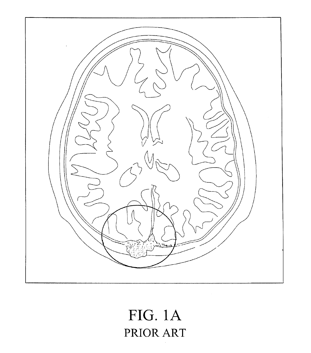
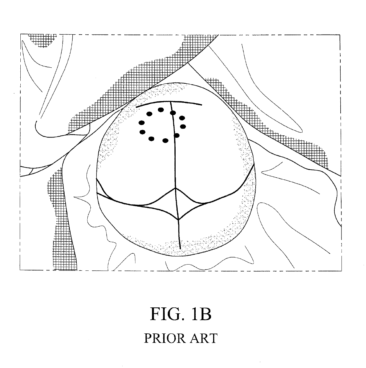
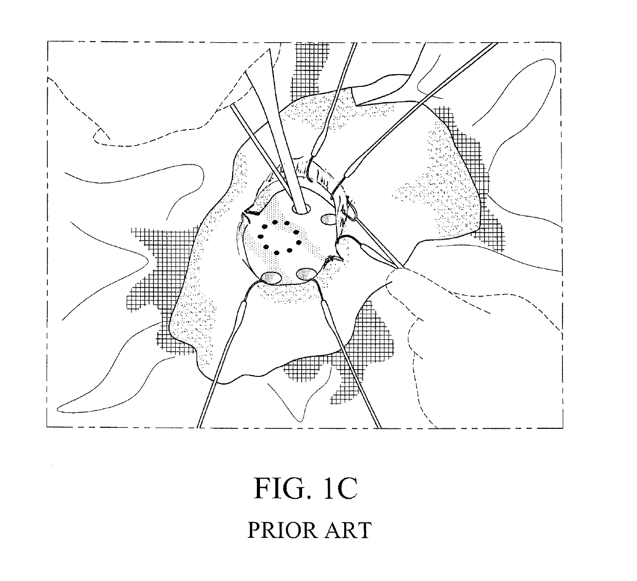
[0053]Referring to FIGS. 3A-3G, 4A, 4B, and 5, the present invention relates to a novel method for performing single-stage cranioplasty with “clear” customized implants (see for example, FIGS. 3C and 3D showing the clear custom craniofacial implant 10 held directly above a skull tumor defect following extirpation (that is, the cranial, craniofacial, and / or facial defect 100 as discussed below) prior to single-stage modification). As explained above in the Background of the Invention, the method of “single-stage cranioplasty” is defined as a surgery where the surgeon intends to create a complica...


PUM


Abstract

A method for performing a cranioplasty includes the steps of prefabricating a sonolucent craniofacial implant based upon information generated by preoperative scans, creating a cranial, craniofacial, and/or facial defect, and attaching the craniofacial implant to the cranial, craniofacial, and/or facial defect. The craniofacial implant is composed of a material that is sonolucent and exhibits attenuation of less than 6 dB/cm.

Description

CROSS REFERENCE TO RELATED APPLICATION[0001]This application is a continuation-in-part of Ser. No. 15 / 957,325, filed Apr. 19, 2018, entitled “METHOD FOR PERFORMING SINGLE-STAGE CRANIOPLASTY RECONSTRUCTION WITH A CLEAR CUSTOM CRANIAL IMPLANT,” which is currently pending, which claims the benefit of U.S. Provisional Patent Application Ser. No. 62 / 489,036, filed Apr. 24, 2017, entitled “METHOD FOR PERFORMING SINGLE-STAGE CRANIOPLASTY RECONSTRUCTION WITH A CLEAR CUSTOM CRANIAL IMPLANT.”BACKGROUND OF THE INVENTION1. Field of the Invention[0002]The present invention generally relates to the field of surgery. In particular, the invention relates to the brain and skull, the art of reconstructing skull defects (i.e., cranioplasty), neurosurgery, neurology, neuroplastic and reconstructive surgery, and craniofacial plastic surgery.2. Description of the Related Art[0003]Craniectomies requiring skull reconstruction (i.e., cranioplasty) are often indicated for a multitude of etiologies including ...

Claims


Application Information

Patent Timeline
IPC IPC(8): A61F2/28A61F2/30
CPCA61F2/2875A61F2/30942A61B2034/105A61B2034/108A61F2002/30948A61F2002/3096A61F2002/3009A61B2017/00924A61B2017/00907A61B17/688A61B17/8061A61F2002/2882
Inventor GORDON, CHAD R.CHRISTOPHER, JESSERABINOVITZ, BRADLEY
Owner LONGEVITI NEURO SOLUTIONS LLC
Why Eureka
Try Eureka