System and method for characterizing drug product impurities

a drug product and impurity detection technology, applied in the field of protein separation methods and cell culture methods, can solve the problems of overestimation of minor lmw species, inability to unambiguously confirm the proposed structure resulting from these methods at the intact protein level, and compromise both drug efficacy and safety, so as to reduce the level of impurities

Inactive Publication Date: 2019-06-27
REGENERON PHARM INC
View PDF0 Cites 0 Cited by
  • Summary
  • Abstract
  • Description
  • Claims
  • Application Information

AI Technical Summary

Benefits of technology

[0007]It is another object of the invention to provide

Problems solved by technology

The formation of HMW species within a therapeutic mAb drug product as a result of protein aggregation can potentially compromise both drug efficacy and safety (e.g. eliciting unwanted immunogenic response) (Rosenberg A S.
However, any proposed structures resulting from these methods cannot be unambiguously confirmed at the intact protein level.
Furthermore, sample preparation conditions employed in SDS-PAGE experiments can generate LMW artifacts through disulfide bond scrambling, which can lead to overestimations of minor LMW species (Zhu Z C, et al.
Unlike SDS-PAGE analysis, these LMW impurities cannot be ext

Method used


Image

  • System and method for characterizing drug product impurities
  • System and method for characterizing drug product impurities
  • System and method for characterizing drug product impurities

Examples


example 1

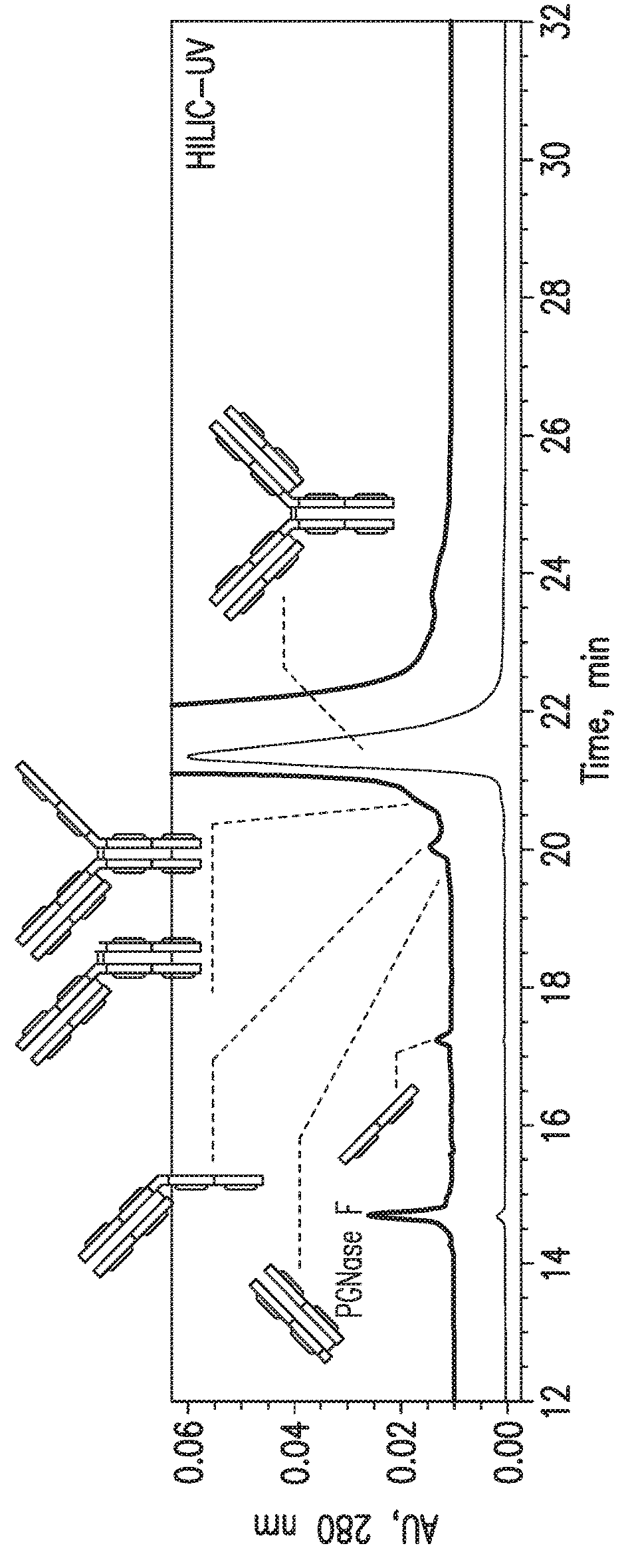
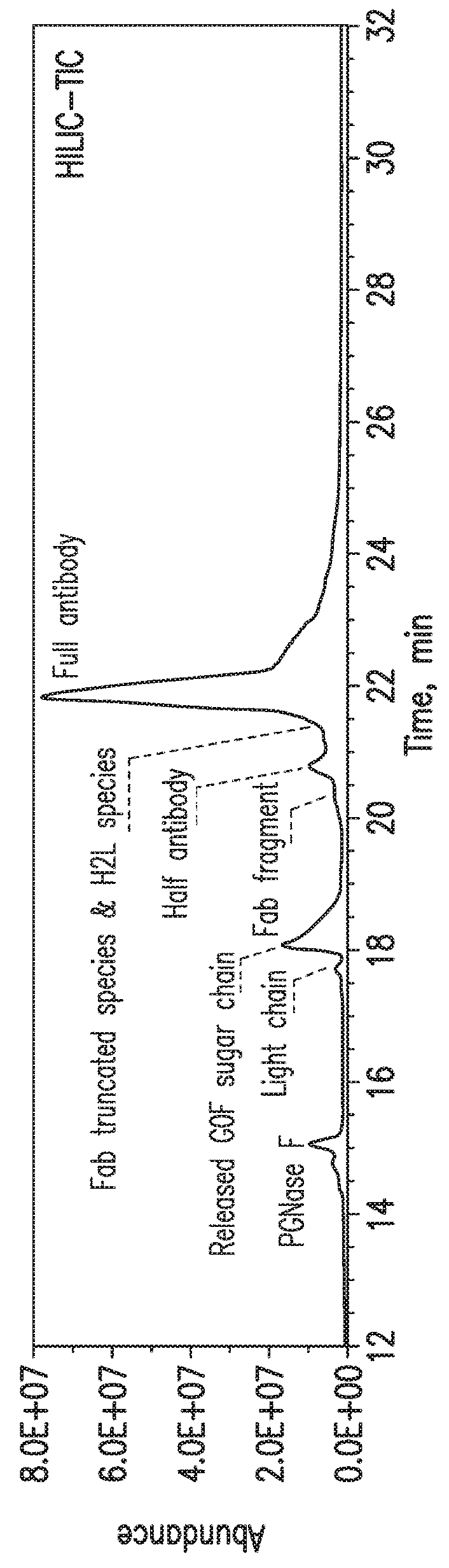
HILIC Separation of mAb-1 Drug Substance Sample

Materials

[0063]For this study, a recombinant IgG1 mAb (mAb-1) made by Regeneron was used. Peptide-N-Glycosidase F (PNGase F, #P0704L) was purchased from New England Biolabs, 1 M Tris-hydrochloride pH 7.5 solution (#15567-027) was purchased from Invitrogen, dithiothreitol (DTT, #20291) was purchased from Thermo Fisher Scientific, and L-cysteine (#168149-25G) was purchased from Sigma-Aldrich. Acetonitrile (LC-MS grade, #A955-4) and trifluoroacetic acid (TFA, #PI28904) were purchased from Fisher Scientific. Milli-Q water was provided in-house.

Methods

[0064]Deglycosylation of mAb-1 and Limited Reduction by DTT and L-Cysteine

[0065]The mAb-1 sample was diluted to a final concentration of 5 μg / μL using 100 mM Tri-HCl (pH 7.5). PNGase F was added at an enzyme to substrate ratio of 1 unit / 10 μg protein. The deglycosylation reaction was conducted at 37° C. for 3 hours. To initiate the limited reduction by DTT, a 20 μg aliquot of the deglycosylated...

example 2

Free Light Chain

Results

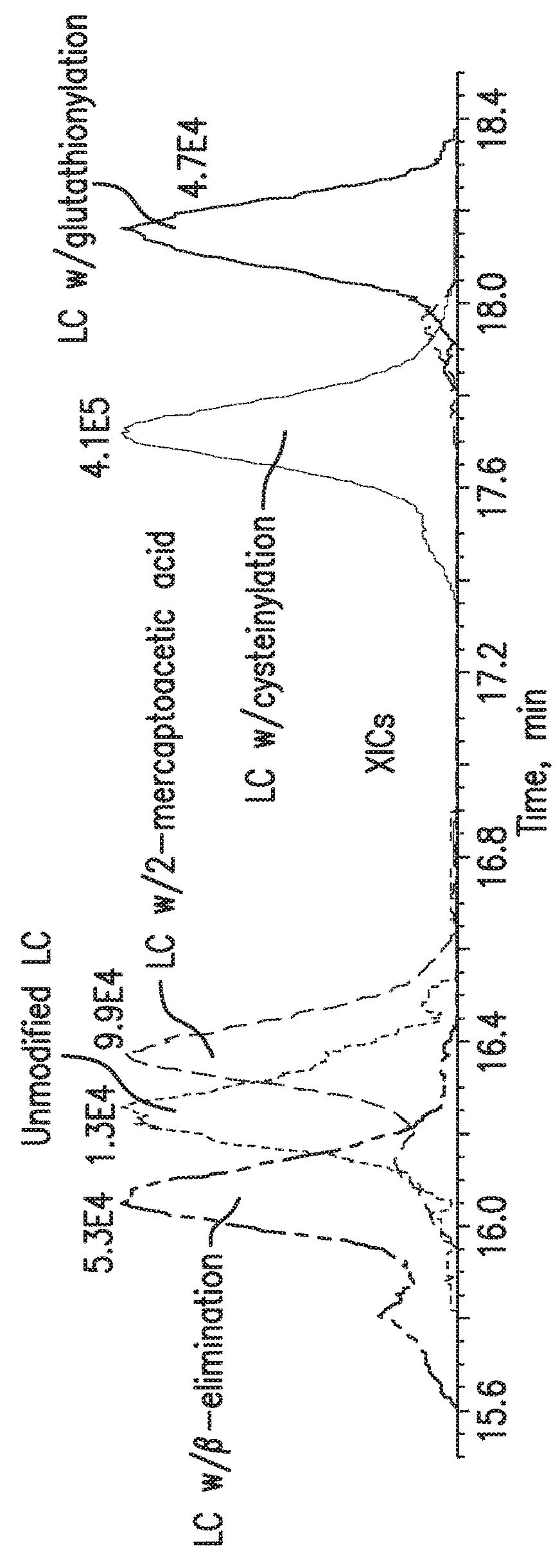
[0069]Multiple light chain-related impurities were detected in HILIC-MS analysis of the mAb-1 sample (FIGS. 2A-2E). The extracted ion chromatograms (XICs) of each species suggested that they also exhibited different retention times during HILIC separation (FIG. 2A). Interestingly, the cysteinylated light chain (+˜119 Da) was identified as the major form of all light chain species present in mAb-1 sample and it is the only species visible by UV (FIG. 2D). The cysteinylation may occur as a result of the thiol-disulfide exchange reaction between the inter-heavy and light chain disulfide bond and a free cysteine molecule, which can be found in cytoplasm. In addition, glutathionylated light chain (+˜306 Da) was also identified, with a retention time slightly later than the cysteinylated light chain (FIG. 2E). Similar to the cysteinylation process, free glutathione (GSH) molecule, which can also be found in cytoplasm, should be responsible for this modification. Ano...

example 3

Fab Fragment

Results

[0070]It is well known that the upper hinge region of heavy chains in an IgG1 molecule is susceptible to hydrolysis leading to the formation of two complementary LMW species, a Fab fragment and a Fab-truncated species (Cordoba A J, Shyong B J, Breen D, Harris R J. Journal of chromatography B: Analytical technologies in the biomedical and life sciences, 818:115-21 (2005). The HILIC-MS method detected both species in the mAb-1 sample. Four major Fab fragments with different masses were identified (FIGS. 3A-3D) and the truncation sites were located by comparing the measured masses with the predicted masses, based on the cDNA-derived amino acid sequence. The amino acid pattern indicated in the deconvoluted mass spectrum matched with the heavy chain hinge region sequence (Cys-Asp-Lys-Thr-His-Thr-Cys). It is worth noting that any Fab fragment should have been readily removed during the purification process due to the lack of the Protein A binding site. Therefore, the pr...


PUM

PropertyMeasurementUnit
Fractionaaaaaaaaaa
Fractionaaaaaaaaaa
Fractionaaaaaaaaaa

Abstract

Systems and methods for characterizing low molecular weight (LMW) protein drug product impurities are provided. One embodiment uses hydrophilic interaction chromatography (HILIC) coupled to mass spectrometry analysis. After removal of the N-linked glycans from the protein drug product, for example an antibody drug product, the elution of LMW impurities from the HILIC column was determined by the size of the molecular weight species. In some embodiments, the HILIC separation is performed under denaturing conditions, making the detection of LMW forms using this method highly comparable to both SDS-PAGE and CE-SDS methods. LMW drug product impurities include, but are not limited to light chain, half antibody, H2L, H2, HL, HC, peptide backbone-truncated species, and combinations thereof.

Description

CROSS REFERENCE TO RELATED APPLICATIONS[0001]This application claims benefit of and priority to U.S. Provisional Patent Application No. 62 / 743,632 filed on Oct. 10, 2018, and U.S. Provisional Patent Application No. 62 / 610,029 filed on Dec. 22, 2017, both of which are incorporated by reference in their entirety.FIELD OF THE INVENTION[0002]The invention is generally directed to protein separation methods and cell culture methods.BACKGROUND OF THE INVENTION[0003]Monoclonal antibodies (mAbs) have been successfully employed to target a wide range of therapeutic areas over the last two decades (Walsh G., Nature biotechnology, 32:992-1000 (2014); Lawrence S. Nature biotechnology, 25:380-2 (2007)). While mAbs possess a conserved covalent heterotetrameric structure consisting of two disulfide-linked heavy chains, each covalently linked through a disulfide bond to a light chain, these proteins often contain low levels of product-related impurities even after extensive purification steps. Low ...

Claims


Application Information

Patent Timeline
IPC IPC(8): C07K16/00G01N33/15
CPCC07K16/00G01N33/15C07K2317/14C07K2317/515C07K2317/55B01D15/305C07K16/065C07K2317/41C07K2317/50G01N2030/027G01N2030/8831C07K2319/00C12Q1/6872G01N33/6848G01N33/6857C12Y305/01052
Inventor WANG, SHUNHAI
Owner REGENERON PHARM INC
Why Eureka
Try Eureka