Method and system for creating and utilizing a patient-specific organ model from ultrasound image data

a patient-specific, ultrasound-based technology, applied in the field of ultrasound imaging, can solve the problems of limited documentation of ultrasound examination, difficult to determine whether a lesion has appeared since a previous examination, and limited value of still image snapshots

Active Publication Date: 2019-07-04
GENERAL ELECTRIC CO
View PDF0 Cites 9 Cited by
  • Summary
  • Abstract
  • Description
  • Claims
  • Application Information

AI Technical Summary

Problems solved by technology

Traditionally, documentation of ultrasound examinations has been limited to still image snapshots of representative structures.
These still image snapshots typically have limited value for re-evaluating an examination or comparing with older examinations.
Still images also make it difficult to determine whether a lesion has appeared since a previous examination.
Such low levels of confidence are incompatible with the demands of diagnostic imaging modalities.
CT and MR imaging modalities, however, are more expensive and invasive than ultrasound imaging.
For example, CT examinations expose patients to radiation and both CT and MR examinations may use a contrast injection that may be harmful to patients with renal challenges.

Method used


Image

  • Method and system for creating and utilizing a patient-specific organ model from ultrasound image data
  • Method and system for creating and utilizing a patient-specific organ model from ultrasound image data
  • Method and system for creating and utilizing a patient-specific organ model from ultrasound image data

Examples


Embodiment Construction

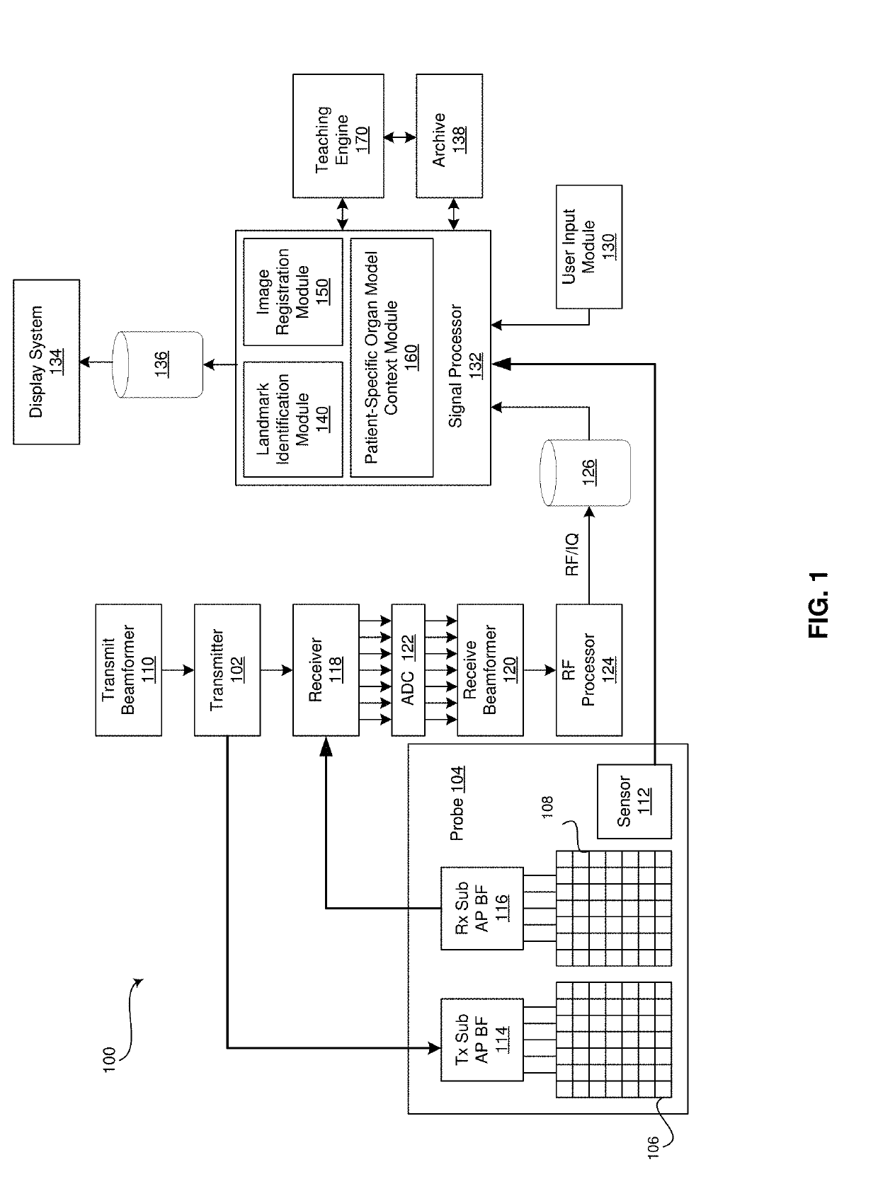
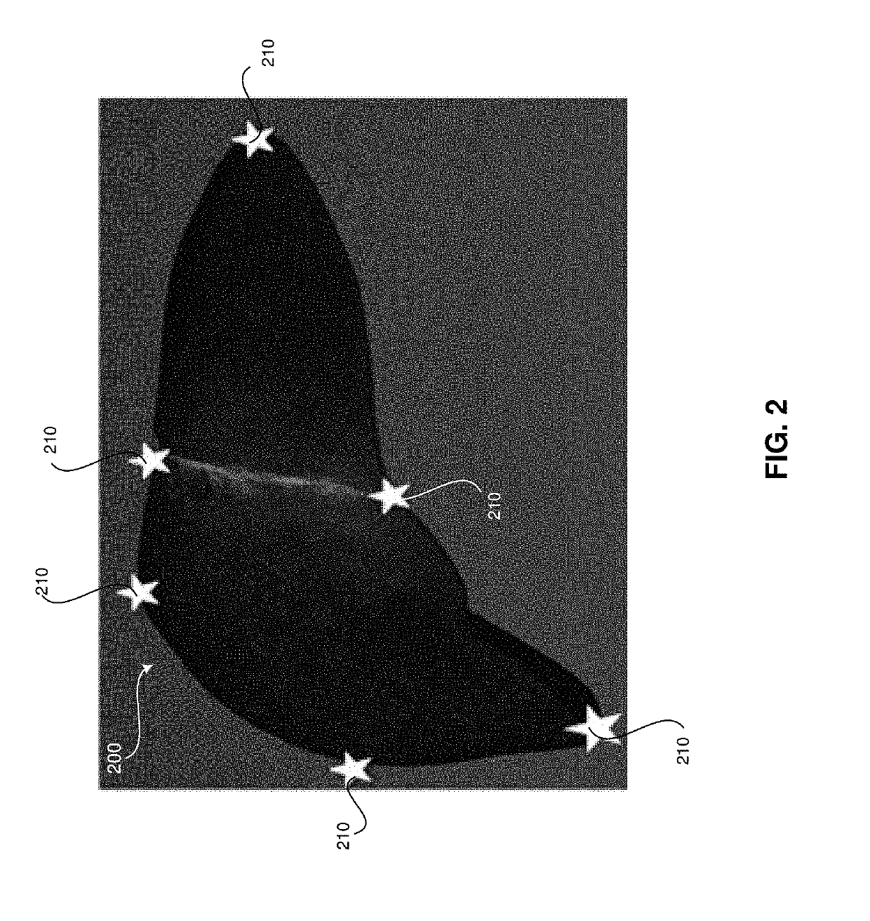
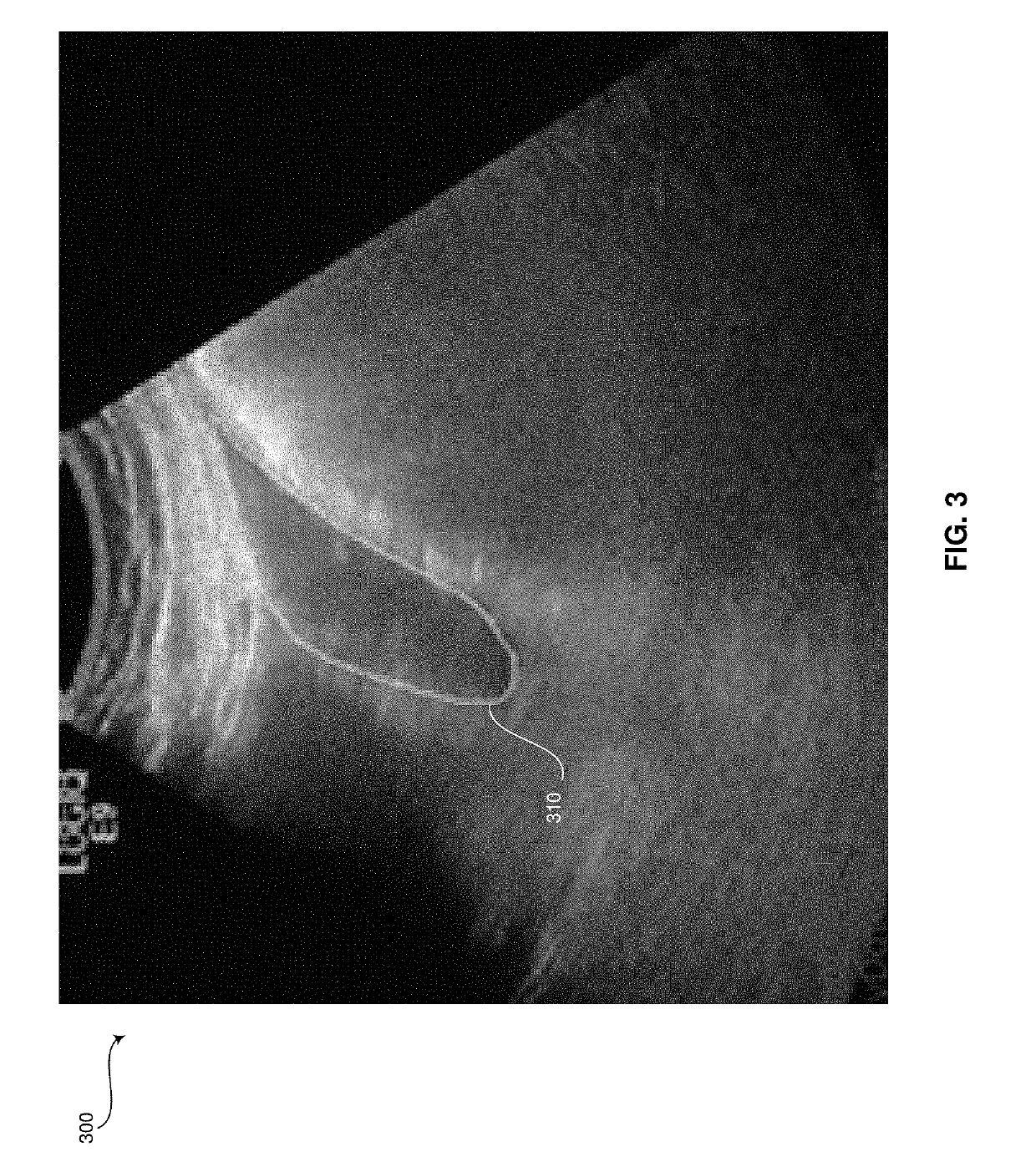
[0018]Certain embodiments may be found in a method and system for generating a patient-specific organ model from ultrasound image data. Various embodiments have the technical effect of providing a three-dimensional context for an associated set of two-dimensional ultrasound image data. The three-dimensional context may include contextual markers illustrating where in a patient-specific organ model a two-dimensional image being viewed is located and / or where in the patient-specific organ model marked structures such as lesions may be found. Moreover, certain embodiments have the technical effect of providing a patient specific model that may be associated with current and / or subsequent two-dimensional ultrasound image data sets based on probe position sensor data associated with the acquired two-dimensional ultrasound image data. Furthermore, various embodiments have the technical effect of providing assistance with real-time navigation of ultrasound-guided procedures by illustrating...


PUM


Abstract

A system and method for generating a patient-specific organ model is provided. The method may include receiving ultrasound images of an organ and probe position data corresponding with each of the ultrasound images. The method may include receiving identification of landmarks in the ultrasound images corresponding with pre-defined landmarks of a generic geometric organ model. The method may include automatically identifying surface points of the organ in the ultrasound images. The method may include generating a patient-specific ultrasound point cloud of the organ based on the received identification of the landmarks, the automatically identified surface points of the organ, and the probe position data. The method may include registering a point cloud of the generic geometric model to the patient-specific ultrasound point cloud to create a patient-specific organ model. The method may include presenting the patient-specific organ model at a display system.

Description

FIELD[0001]Certain embodiments relate to ultrasound imaging. More specifically, certain embodiments relate to a method and system for generating a patient-specific organ model from ultrasound image data. The patient-specific organ model may be utilized in ultrasound-guided procedures, follow-up studies, and / or to otherwise provide a three-dimensional context to a set of two-dimensional ultrasound images.BACKGROUND[0002]Ultrasound imaging is a medical imaging technique for imaging organs and soft tissues in a human body. Ultrasound imaging uses real time, non-invasive high frequency sound waves to produce a two-dimensional (2D) image and / or a three-dimensional (3D) image.[0003]Traditionally, documentation of ultrasound examinations has been limited to still image snapshots of representative structures. These still image snapshots typically have limited value for re-evaluating an examination or comparing with older examinations. Still images also make it difficult to determine whether...

Claims


Application Information

Patent Timeline
Patent Type & Authority Applications(United States)
IPC IPC(8): A61B8/08A61B8/00G06T7/00
CPCA61B8/5246A61B8/5207A61B8/5223A61B8/461A61B8/4245G06T7/0014G06T2207/10136G06T2207/30204G06T2207/10028G06T2207/20081G06T2207/30096G06T2207/20101A61B8/0833A61B8/085A61B8/4254A61B8/4488G06T7/12G06T7/149G06T2207/10132G06T2207/30056G16H50/30
Inventor SUDHAKAR, PRASADLANNING, JUSTIN DANIELANNANGI, PAVAN KUMARWASHBURN, MICHAEL
Owner GENERAL ELECTRIC CO
Why Eureka
Try Eureka