Systems and method for x-ray imaging

a fluoroscopic imaging and x-ray technology, applied in the field of medical imaging, can solve the problems of patient exposure to more radiation than desired, and the difficulty of obtaining high-quality images,

Active Publication Date: 2019-08-15
GENERAL ELECTRIC CO
View PDF5 Cites 4 Cited by
  • Summary
  • Abstract
  • Description
  • Claims
  • Application Information

AI Technical Summary

Benefits of technology

[0005]In one embodiment, a method for an x-ray imaging system includes acquiring, with the x-ray imaging system, a plurality of images as an x-ray tube current of the x-ray imaging system is ramping from a predefined x-ray tube current to an updated x-ray tube current, the updated x-ray tube current determined based on an estimated patient thickness estimated from a prior image acquired with the x-ray imaging system while the x-ray tube current is at the predefined x-ray tube current. The method further includes combining the plurality of images into a final image and outputting the final image for display via a display device. In this way, patient thickness may be estimated during an initial phase of an imaging session, based on an image acquired during the imaging session, and used to command the x-ray imaging system to an updated x-ray current that is based on the patient thickness. As the x-ray tube current is ramping toward the updated x-ray tube current, a plurality of images is acquired and combined to generate a final image for display. By doing so, a final image of sufficient quality to view the underlying patient anatomy may be generated in a short amount of time, e.g., while the current is changing, rather than waiting until the current has reached the updated current.

Problems solved by technology

Due to the short exposure time, obtaining a high-quality image may be problematic and / or the patient may be subject to more radiation than desired.

Method used


Image

  • Systems and method for x-ray imaging
  • Systems and method for x-ray imaging
  • Systems and method for x-ray imaging

Examples


Embodiment Construction

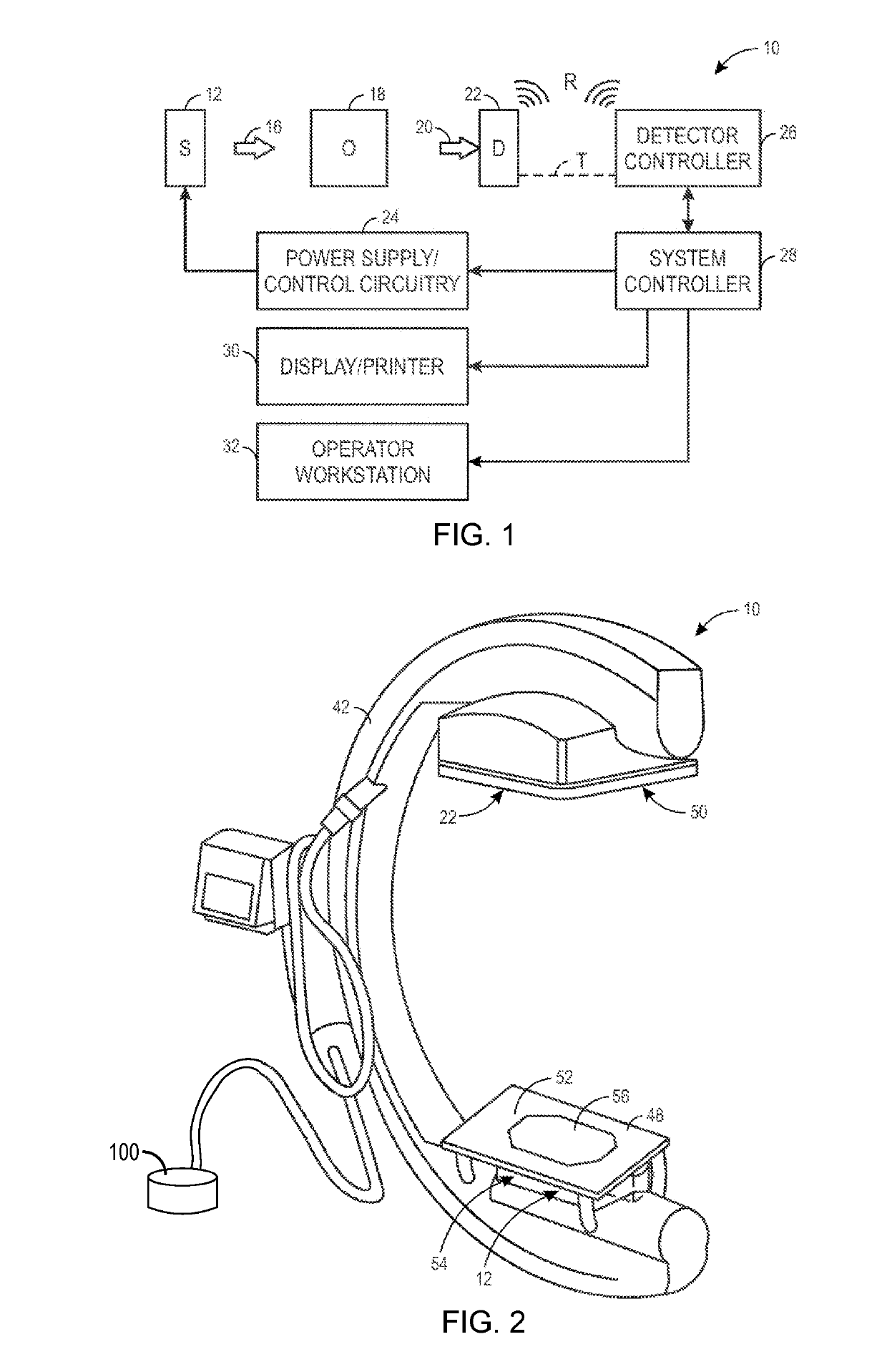
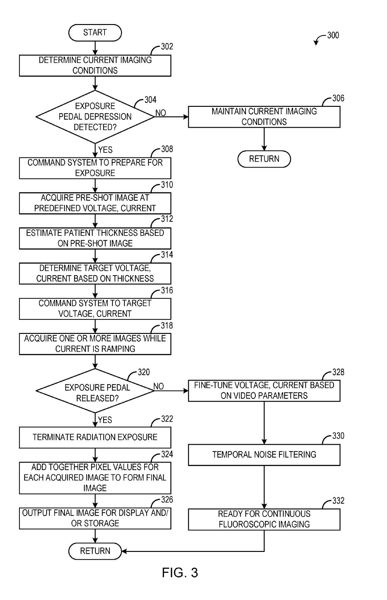
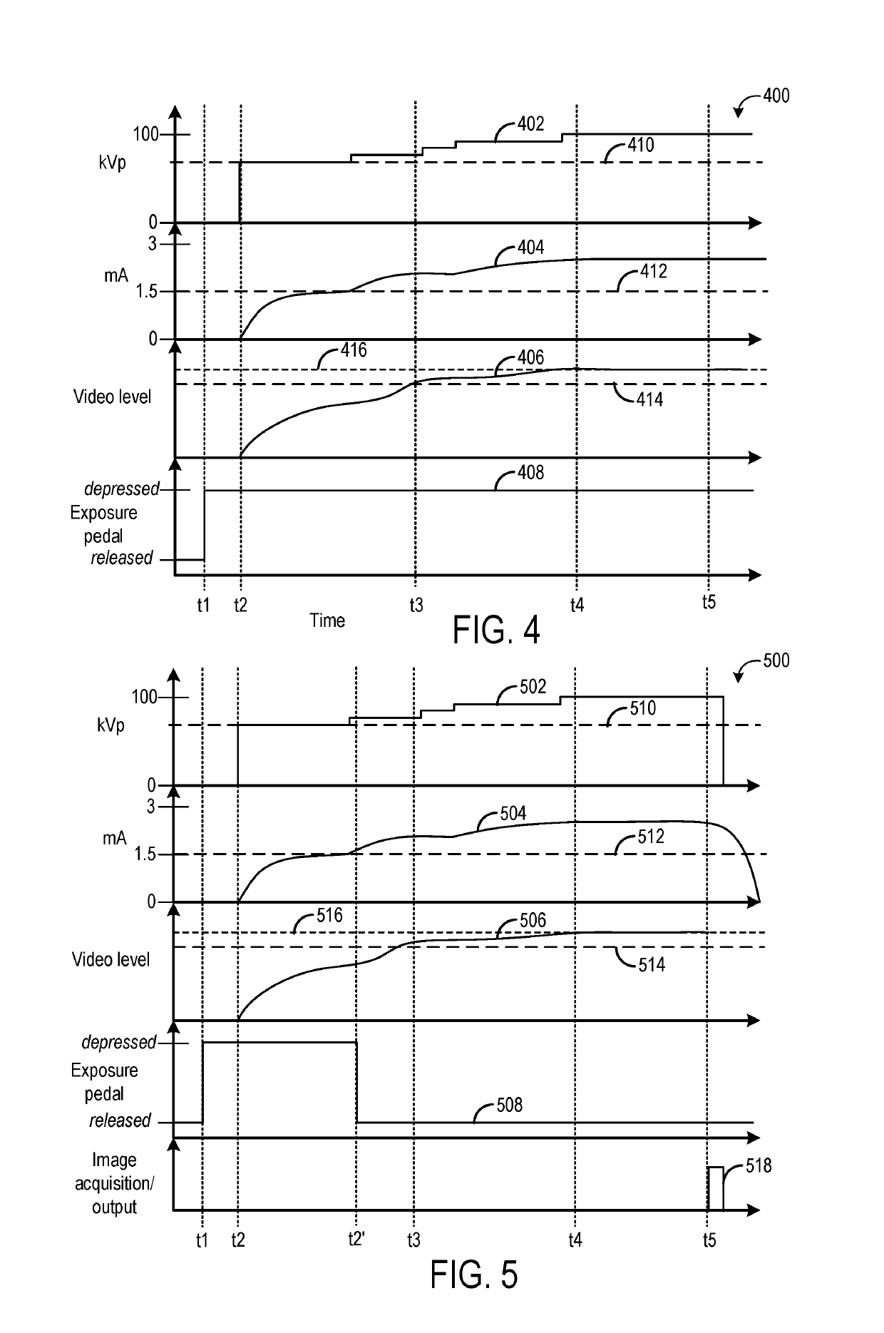
[0013]The following description relates to various embodiments of reducing exposure time during acquisition of a single skeletal x-ray image. During a surgical procedure monitored / assisted by fluoroscopic imaging, continuous, real-time x-ray images of a patient may be displayed, allowing clinicians to monitor movement of anatomical features. During such procedures, it may also be useful to occasionally monitor the patient using single, still x-ray images. To initiate fluoroscopic imaging, an exposure control pedal may be depressed, e.g., by a foot of a clinician. A quick depression and release (e.g., tap) of the exposure control pedal may indicate the clinician is requesting a single x-ray image, where prolonged depression of the exposure control pedal may indicate the clinician is requesting fluoroscopic imaging.

[0014]Typically, to obtain a high-quality x-ray image, a sequence of steps are performed before a final image is acquired and output for display. These steps may include pr...


PUM


Abstract

Methods and systems are provided for controlling an x-ray imaging system. In one embodiment, a method for an x-ray imaging system, includes acquiring, with the x-ray imaging system, a plurality of images as an x-ray tube current of the x-ray imaging system is ramping from a predefined x-ray tube current to an updated x-ray tube current, the updated x-ray tube current determined based on an estimated patient thickness estimated from a prior image acquired with the x-ray imaging system while the x-ray tube current is at the predefined x-ray tube current, combining the plurality of images into a final image, and outputting the final image for display via a display device.

Description

FIELD[0001]Embodiments of the subject matter disclosed herein relate to medical imaging, and more particularly, to x-ray fluoroscopic imaging.BACKGROUND[0002]Non-invasive imaging technologies allow images of the internal structures or features of a patient or object to be obtained without performing an invasive procedure on the patient or object. In particular, such non-invasive imaging technologies rely on various physical principles, such as the differential transmission of x-rays through the target volume or the reflection of acoustic waves, to acquire data and to construct images or otherwise represent the observed internal features of the patient or object.[0003]For example, in fluoroscopy and other x-ray based imaging technologies, x-ray radiation is directed toward a subject, typically a patient in a medical diagnostic application, a package or baggage in a security screening application, or a fabricated component in an industrial quality control or inspection application. A ...

Claims


Application Information

Patent Timeline
Patent Type & Authority Applications(United States)
IPC IPC(8): A61B6/00G01B15/02
CPCA61B6/482A61B6/487A61B6/5235A61B6/544A61B6/463A61B6/405A61B6/42A61B6/4441G01B15/02G01B15/025
Inventor LIU, JAMES ZHENGSHECHANDRA, NAVEEN STEPHAN
Owner GENERAL ELECTRIC CO
Why Eureka
Try Eureka