
Acpa-negative ra diagnostic marker and application thereof

a diagnostic marker and ra technology, applied in the field of biological detection, can solve the problems of low sensitivity to ra diagnosis, difficult to provide accurate diagnosis and treatment, and difficult to be detected

Inactive Publication Date: 2019-12-05
PEKING UNION MEDICAL COLLEGE HOSPITAL CHINESE ACAD OF MEDICAL SCI
View PDF0 Cites 0 Cited by
  • Summary
  • Abstract
  • Description
  • Claims
  • Application Information

AI Technical Summary

Benefits of technology

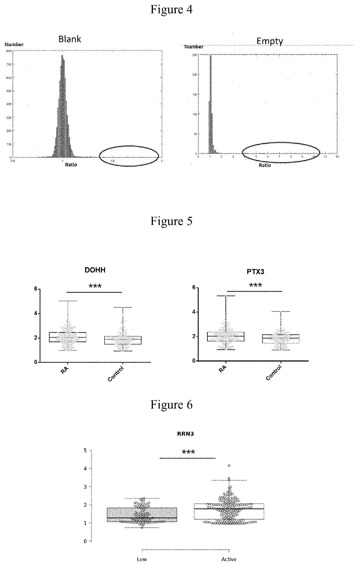
The present invention identified a set of autoantigens that can be used as diagnostic markers for rheumatoid arthritis (RA). These autoantigens have a high sensitivity and specificity, with some able to predict disease activity and therapeutic efficacy. The results were validated using a protein chip containing a larger set of RA autoantigens, which successfully distinguished between RA and other autoimmune diseases, and between different levels of disease activity. The identification of these autoantigens can contribute to the development of more effective diagnostic and therapeutic tools for RA.

Problems solved by technology

However, too strict definition for arthritis in the classification criteria leads to a low sensitivity to RA diagnosis in practice, a large number of early RA patients failed to be identified.
The lack of specific biomarkers for ACPA-negative RA patients makes it quite difficult to provide accurate diagnosis and treatment.
Although these two antibodies have high specificity in the diagnosis of RA, they are difficult to be detected.
Some patients' symptoms are self-limiting and mild, while some suffer from rapidly progressive inflammation, joint destruction and severe disability.
The heterogeneity of RA clinical manifestations leads to great differences of reactions to treatment.
For now, there's no way to predict the effect of specific treatment for the lack of efficient biomarkers to sub-classify RA patients.
Many ACPA-negative RA patients have limited targets to subclassify, because the lack of potent biomarkers leads to limited targets to classify the clinical manifestation of RA.

Method used


Image

  • Acpa-negative ra diagnostic marker and application thereof
  • Acpa-negative ra diagnostic marker and application thereof
  • Acpa-negative ra diagnostic marker and application thereof

Examples


example 1

Identification of Candidate RA Autoantigens Using High-Density Protein Chips

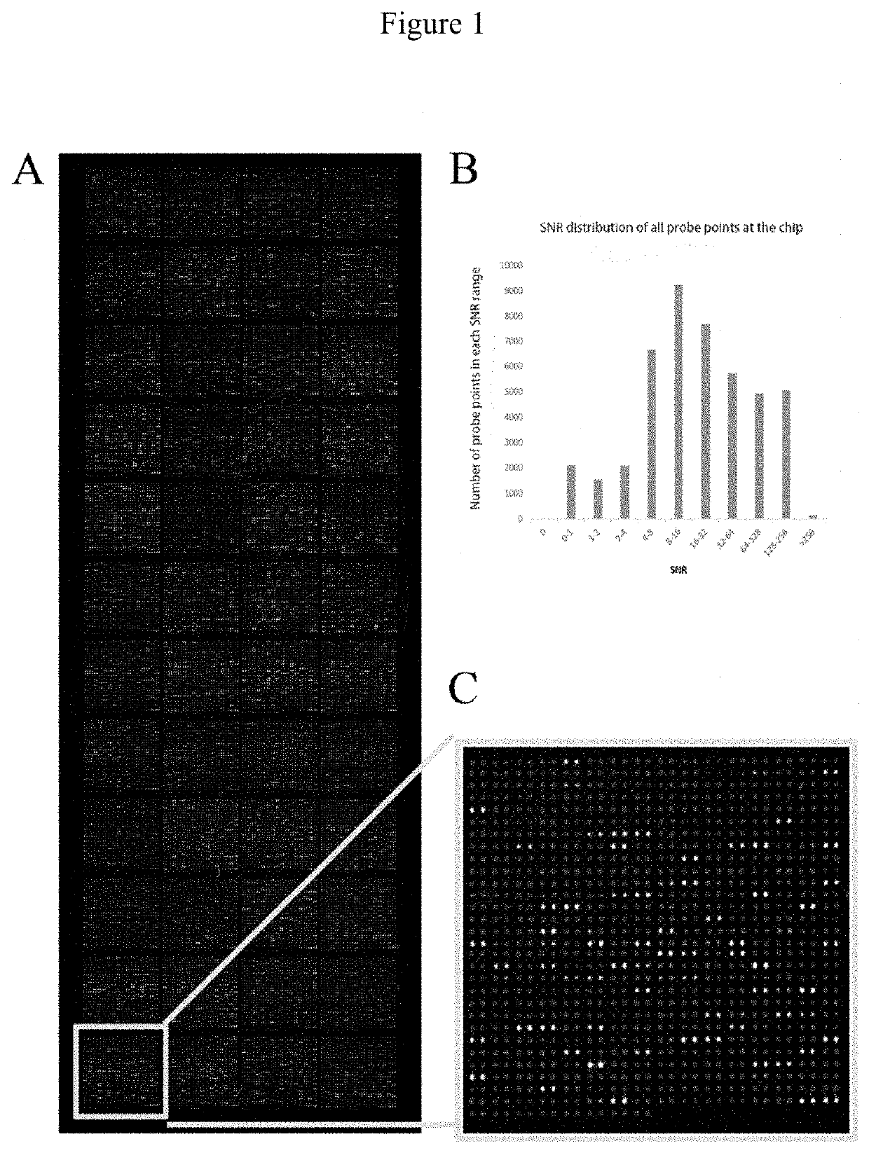
[0053]The high-density protein chips and Saccharomyces cerevisiae-expressing recombinant vectors including target gene sequences were provided by Dr. Zhu's laboratory at Johns Hopkins University. Each chip consisted of 48 blocks and the block included 992 probe points arranged in a 32*31 array, with 2 parallel points for each protein probe. The protein chip consisted of 21827 non-redundant recombinant human proteins. The recombinant proteins, with glutathione S-transferase (GST) tag at the N-terminus, were derived from the full-length open reading frame (ORF) of the corresponding gene expressed by Saccharomyces cerevisiae.

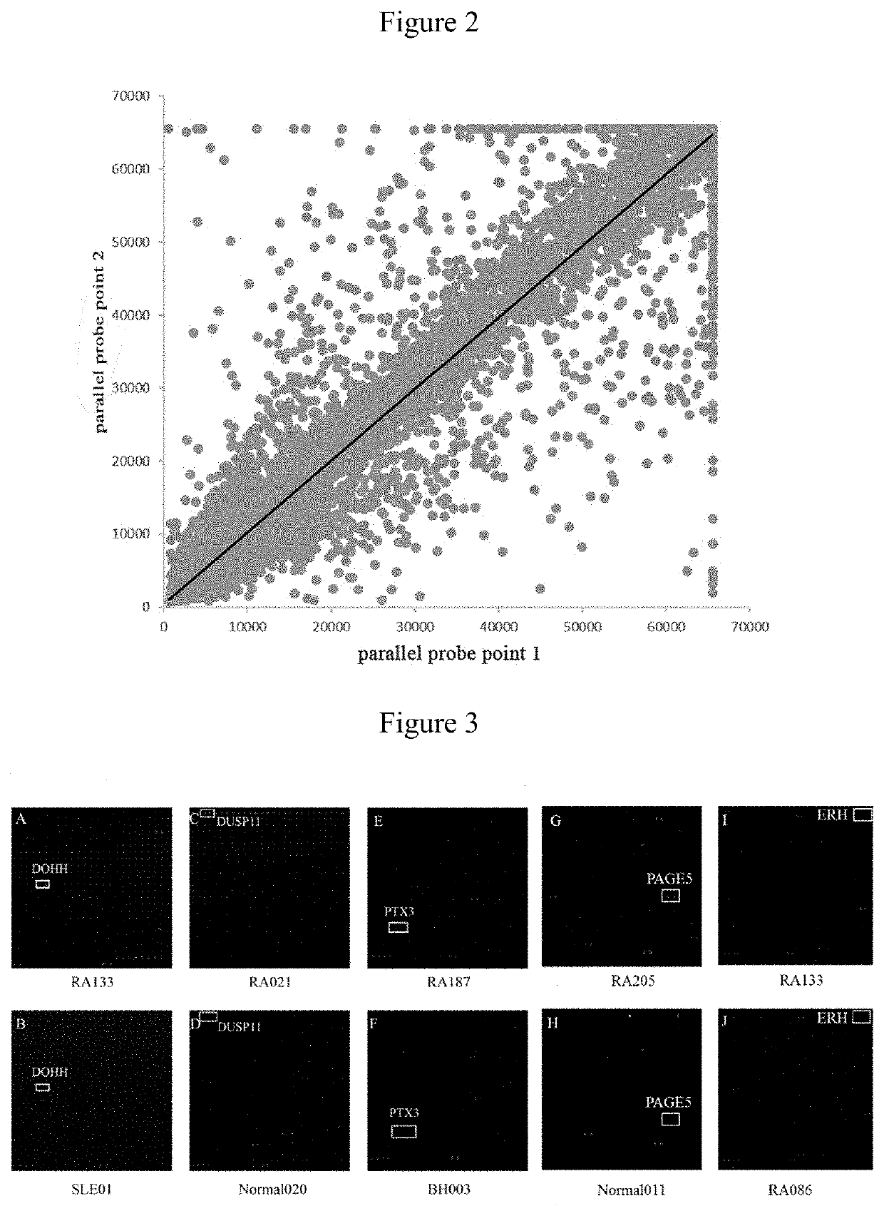
[0054]The quality of chips was verified by hybridizing mouse anti-GST monoclonal antibodies with the chips. Qualified repeatability of duplicate protein spots was achieved when the correlation coefficient of fluorescent signal value between duplicate spots reached 97%.

[0055]Each high-densi...

example 2

Hybridization of High-Density Protein Chips, RA and Control Serum

[0056]60 RA and 60 control (10 BD, 10 TA, 10 SLE and 30 healthy controls) serum samples were hybridized with 120 protein chips, and candidate RA autoantigens were identified by signal collection and data analysis. PE-Cy5 labeled anti-human IgG antibody was used to detect the reaction between the serum autoantibodies and autoantigen probes. FIG. 3 shows the representative partial scan image formed by serum hybridization with high-density protein chips, different protein antigen probe is shown in the box. A, C, E, G show scan images of hybridization of 4 RA serum samples with chips. B, D, F, H show scan images of hybridization of 4 control serum samples (including disease and healthy controls) with chips. FIG. 1 shows the scan image of RA treatment effective. Figure J shows the scan image of RA treatment ineffective. Two parallel points protein probes in the boxes of Figures A and B are DOHH, DUSP11 in the boxes of Figur...

example 3

Construction of RA Autoantigen Protein Chip and Verification of Serum Screening

[0062]By analyzing the result of hybridizing small number of serum samples with high-density protein chips, 46 candidate RA autoantigens were identified. To verify the specificity and sensitivity of these autoantigens, the present invention constructed low probe density RA autoantigen protein chip. Table 2 shows the distribution of microarray of each probe at RA autoantigens protein chip. The probes at the chip included 46 candidate RA autoantigens and 5 control probes (IGHG1).

TABLE 2Microarray of each probe at RA autoantigens protein chipAK2IGHG1NDATP13A5NDTBC1D19RAB35UBXN10RAB3DAPH1ATNFAIP1HDAC4ARL2BPRAI14RRN3POLR3BERHNDRG1BLANKBLANKBLANKBLANKBLANKGARSSUGT1IGHG1NOL3ZSCAN20LSP1RGCCEMPTYPAGE5FGF12FAM84ADOHHNECAB1NDEL1DUSP11PDCD2MYLKSTK24METTL21CIGHG1STK3BABAM1DGKKPTX3PPFIA4EMPTYSPANXN2IGHG1CHAC2RNF183ATXN10IGHG1EMPTYCHST11PLEKHG2SNX33BLANK

[0063]51 probes at RA autoantigen protein chip all had duplicate po...


PUM


Abstract

The invention provides a use of a deoxyhypusine dioxygenase DOHH or a fragment thereof in the preparation of a reagent for diagnosing an anti-citrulline polypeptide antibody-negative rheumatoid arthritis disease. 35 proteins as candidate ACPA-negative RA autoantigens were screened by hybridizing high density protein chips with RA serum. 4 protein antigens (DOHH, DUSP11, PTX3 and PAGE5) were identified as having high sensitivity and specificity in the ACPA-negative serum of RA, wherein DOHH can be used as the ACPA-negative RA diagnostic markers.

Description

TECHNICAL FIELD[0001]The present invention belongs to biological detection field, in particular, relates to a kind of ACPA-negative RA diagnostic marker and application thereof.BACKGROUND ART[0002]Rheumatoid arthritis (RA) is a chronic autoimmune disease, mainly characterized by the multiple joint inflammation and local bone destruction. In developing countries, rheumatoid arthritis affects nearly 0.5% to 1% of the population. In general, the incidence of RA in women is higher than that in men, and the elderly are more likely to develop RA than the youth. The clinical manifestations of rheumatoid arthritis display heterogeneity, ranging from self-limiting disease with mild symptoms to fast developed inflammation along with joint destruction and severe physical disability. Due to differences in disease performance, classification criteria were developed as the basis for disease definition, selection of standardized clinical trials, and comparison of multicenter studies. In 1987, the ...

Claims


Application Information

Patent Timeline
IPC IPC(8): G01N33/68G01N33/564
CPCG01N2800/102G01N33/564G01N33/6893G01N33/573G01N2333/90241
Inventor ZHANG, XUANMO, WENXIULI, YONGZHEHU, CHAOJUNLIU, GUOZHENWU, LIJUN
Owner PEKING UNION MEDICAL COLLEGE HOSPITAL CHINESE ACAD OF MEDICAL SCI
Learn More