Methods for Determining Contrast Agent Concentration Using Magnetic Resonance Imaging

a magnetic resonance imaging and contrast agent technology, applied in the direction of nmr measurement, instruments, applications, etc., can solve the problems of introducing errors and often both very rapid rates

Active Publication Date: 2020-03-05
MAYO FOUND FOR MEDICAL EDUCATION & RES
View PDF0 Cites 1 Cited by
  • Summary
  • Abstract
  • Description
  • Claims
  • Application Information

AI Technical Summary

Problems solved by technology

For malignancies both of these rates are often both very rapid.
It is possible to apply multiple pulse sequences to determine T10 or to assume some typical, population-based value, but these values may introduce error as they are not patient specific.

Method used


Image

  • Methods for Determining Contrast Agent Concentration Using Magnetic Resonance Imaging
  • Methods for Determining Contrast Agent Concentration Using Magnetic Resonance Imaging
  • Methods for Determining Contrast Agent Concentration Using Magnetic Resonance Imaging

Examples


Embodiment Construction

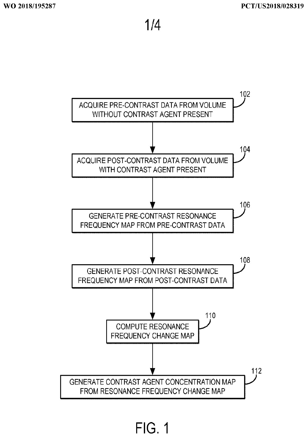
[0023]Described here are methods for measuring or otherwise determining the concentration (e.g., the absolute concentration) of a contrast agent using magnetic resonance imaging (“MRI”). As stated above, contrast agents used in MRI typically change the relaxation rate of the spins that are proximate to the contrast agent. As one non-limiting example, the contrast agent can be a gadolinium (“Gd”) based contrast agent, which increases the longitudinal relaxation rate (R1), or decreases the longitudinal relaxation time (T1), of the surrounding spins, which may be hydrogen nuclei or other spins from which magnetic resonance signals are to be obtained. In other examples, the contrast agent can include other paramagnetic contrast agents (e.g., manganese based agents), superparamagnetic materials (e.g., superparamagnetic iron oxide based agents), or other contrast agents that affect the longitudinal relaxation, transverse relaxation, or both, of nuclear spins.

[0024]Rather than estimate the...


PUM


Abstract

The present disclosure provides systems and methods for measuring a concentration of a contrast agent using magnetic resonance imaging (MRI). The method includes acquiring pre- and post-contrast data from a volume of a subject, where the pre-contrast data is acquired before a contrast agent is administered and the post-contrast data is acquired after the contrast agent is administered. A pre- and post-contrast resonance frequency map are then computed, where the pre-contrast frequency map is based on one or more pre-contrast resonance frequency values, and the post-contrast resonance frequency map is based on one or more post-contrast resonance frequency values. The pre- and post-contrast frequencies maps are then used to generate a resonance frequency change map that is subsequently used to generate a contrast agent concentration map that indicate a concentration of the contrast agent that was present at each voxel in the volume at the second time.

Description

CROSS-REFERENCE TO RELATED APPLICATIONS[0001]This application claims the benefit of U.S. Provisional Patent Application Ser. No. 62 / 487,845, filed on Apr. 20, 2017, and entitled “GADOLINIUM-INDUCED SUSCEPTIBILITY EFFECTS IN SINGLE-ECHO DIXON CE-MRA,” which is herein incorporated by reference in its entirety.STATEMENT REGARDING FEDERALLY SPONSORED RESEARCH[0002]This invention was made with government support under EB000212 awarded by the National Institutes of Health. The government has certain rights in the invention.BACKGROUND[0003]Contrast agents are used in many imaging modalities for a variety of purposes. They are generally administered in some fashion and then selectively flow to or are taken up by specific tissues, organs, or cell types within the body. Sometime after administration the distribution of the contrast agent is imaged using a specific modality such as nuclear medicine, computed tomography (CT), or magnetic resonance imaging (MRI). As one example, contrast agents ...

Claims


Application Information

Patent Timeline
Patent Type & Authority Applications(United States)
IPC IPC(8): A61B5/055G01R33/56G01R33/48G01R33/485
CPCG01R33/485G01R33/5601G01R33/5608G01R33/4828A61B5/055
Inventor RIEDERER, STEPHEN J.STINSON, ERIC G.
Owner MAYO FOUND FOR MEDICAL EDUCATION & RES
Why Eureka
Try Eureka